Gram Staining: Definition, Procedure, Facts, Topics, Diagram
Gram staining is a differential staining method that has been in common use for classifying bacteria into Gram-positive and Gram-negative groups according to cell wall type. Its use has been boosted in recent times through the combination of its use with automated digital microscopy and artificial intelligence-based image analysis to accelerate pathogen identification, particularly in life-threatening infections such as sepsis (Journal of Clinical Microbiology, 2023). A study in Nature Microbiology (2022) further explains that differences in peptidoglycan structure between bacterial species may influence staining results, pointing to the importance of cell wall biology in clinical microbiology. Improvements in microfluidic technology also optimised Gram staining by fast, on-chip staining and analysis of bacterial samples to significantly decrease turnaround time in clinical practice (Lab on a Chip, 2023).
This Story also Contains
- What Is Gram Staining?
- Principles Of Gram Staining
- Importance Of Gram Staining
- Recommended video for "Gram Staining"

In addition, scientists are currently investigating quantitative Gram staining techniques that quantify dye retention more accurately, making semi-automated diagnosis with increased reproducibility possible. Research also indicates that antibiotic resistance mechanisms may change cell wall composition at times, possibly affecting Gram reaction outcomes (Frontiers in Microbiology, 2022). Such advancements support the continued applicability and versatility of Gram staining in contemporary microbial diagnostics and research.
What Is Gram Staining?
In microbiology, the Gram stain or Gram’s method is a method used for distinguishing bacteria based on the characteristics of the cell wall. Invented in 1884 by Danish scientist Dr Hans Christian Gram, it helps the use of crystal violet dye, iodine, alcohol and safranin. Gram staining is used principally to distinguish bacteria into two groups, namely the Gram-positive and the Gram-negative. Septum thickness is thicker in Gram-positive bacteria compared to Gram-negative bacteria. Gram-positive bacteria retain the violet stain because of the thick layer of peptidoglycan, while Gram-negative bacteria lose the stain due to the thin layer of peptidoglycan and a membranous layer surrounding it.
Also Read
Principles Of Gram Staining
Gram staining is a differential staining method invented by Hans Christian Gram to categorise bacteria into two broad categories: Gram-positive and Gram-negative. The mechanism relies on the variation in composition and thickness of the bacterial cell wall, more specifically, the peptidoglycan layer. The crystal violet stain forms an iodine complex, which becomes entrapped within the thick peptidoglycan layer of Gram-positive bacteria, causing them to stain purple. Conversely, Gram-negative bacteria, with a thinner peptidoglycan layer and an outer membrane, lose the crystal violet-iodine complex upon decolourisation and incorporate the counterstain (safranin) and become pink. Some of the basic points about Gram staining are discussed below:
- According to the cell wall structure, particularly the peptidoglycan thickness
- Gram-positive bacteria hold the crystal violet because they have thick peptidoglycan
- Gram-negative bacteria lose the main stain and take up safranin
- Helps in bacterial classification and directs antibiotic therapy
Cell Wall Composition
The structure of the bacterial cell wall determines the Gram reaction upon staining. It gives rigidity and protection to the cell. According to the structure of their cell walls, bacteria are divided into Gram-positive and Gram-negative bacteria. This is important in microbiology since it affects the staining results and the response of the bacteria to antibiotics.
Comparison of Cell Wall Composition: Gram-Positive vs Gram-Negative Bacteria
The basic comparison between the cell wall composition of Gram-positive and Gram - Gram-negative bacteria is discussed below:
| Feature | Gram-Positive Bacteria | Gram-Negative Bacteria |
|---|---|---|
| Peptidoglycan Layer | Thick, multilayered | Thin, single layer |
| Outer Membrane | Absent | Present, composed of lipopolysaccharides |
| Teichoic Acids | Present | Absent |
| Stain Retention | Retains crystal violet → appears purple | Loses crystal violet, retains safranin → appears pink |
| Sensitivity to Antibiotics | More sensitive to antibiotics like penicillin | Less sensitive due to protective outer membrane |
| Overall Cell Wall Structure | Simple | Complex |
Structural differences in Peptidoglycan layers
The purification of gram-positive bacteria has a resistant and homogeneous layer of peptidoglycan that retains the crystal violetiodine complex, explaining why they take the purple stain. However, due to the thin layer of peptidoglycan in the middle of the outer and inner membranes present in the gram-negative bacteria, the crystal violet stain is decolourised during the decolourisation step, and the safranin counter stain is then taken up, hence appearing pink or red.
Staining Procedure
The staining technique is a group of steps to stain microbial cells for visibility and differentiation under a microscope. It helps treat fixed bacterial smears on a slide with particular dyes. It focuses on structural variations in cell walls, which facilitate bacterium classification. Gram staining is among the most universal differential staining methods in microbiology.
Step-by-step process:
| Step | Description |
|---|---|
| Fixation | Heat fixation is done by heating the bacterial smear, causing bacteria to firmly attach to the slide. |
| Crystal Violet Staining | Slide is flooded with crystal violet, a primary stain that colours all cells purple for better visibility. |
| Iodine Treatment | Iodine solution is applied to form a complex with crystal violet inside the cells, increasing stain retention. |
| Alcohol Decolourisation | Alcohol or ethanol wash removes stain from cells that fail to retain it, making them colourless. |
| Counterstaining with Safranin | Slide is stained with safranin, dyeing decolourised cells pink to contrast with other stained cells, helping differentiation. |
Interpretation of Results
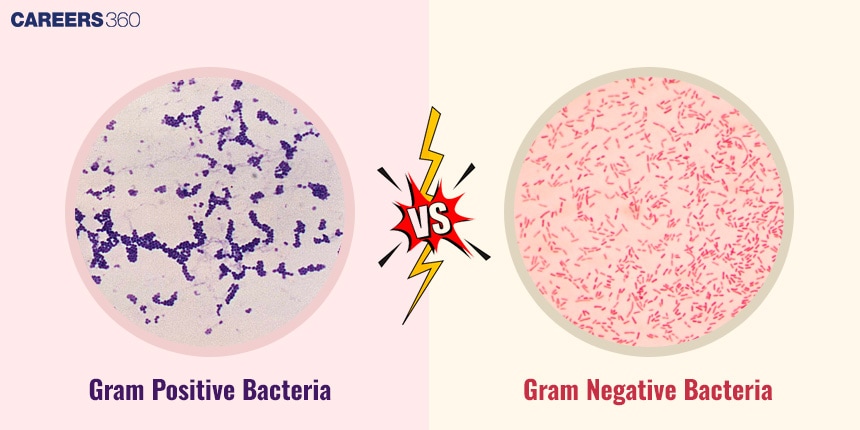
The Gram stain result is interpreted based on the colour of the bacterial cells under a microscope. Gram-positive bacteria appear purple due to crystal violet retention, while Gram-negative bacteria appear pink after taking up the safranin counterstain.
Gram-positive bacteria:
Such bacteria stain purple or blue when viewed under the microscope because they can retain the crystal violet stain. This is because they have a thick layer of peptidoglycan in their cell walls, trapping the dye even after decolourisation. Some important facts are discussed below:
- Have a thick layer of peptidoglycan
- Retain the crystal violet stain when decolourised
- Are purple or blue
Gram-negative bacteria:
They stain pink or red following Gram staining. They lose the stain of crystal violet during decolourisation due to their thin peptidoglycan layer and the fact that they contain a further outer membrane. They do retain safranin counterstain, giving them their distinctive colour. Some important facts are discussed below:
- Contain a thin peptidoglycan layer and an outer membrane
- Loss of stain of crystal violet during decolourisation
- Keep safranin counterstain
- Look pink or red under the microscope
Diagram of Gram-positive and Gram-negative bacteria post-staining.
Importance Of Gram Staining
Gram staining is an important microbiological technique with broad application areas in clinical, research, and epidemiological fields. It is a rapid and inexpensive technique to distinguish between bacteria according to the morphology of their cell wall, and this has important implications for therapy and analysis.
Clinical Applications in the Diagnosis Of Bacterial Infections
In clinics, Gram staining plays a significant role in the proper identification of Gram-positive and negative bacteria and the choice of antibiotics to the characteristics of bacteria found in a sample.
Role In Research, Epidemiology, And Antibiotic Susceptibility Testing
In research, this staining technique helps the classifiers in the identification of bacteria, in particular their morphology and how they are pathogenic. It is also used in epidemiology, for example, in revealing the behaviour of bacterial infections in a given population and in antibiotic susceptibility testing, where one is bound to determine the efficiency of the antibiotics against different types of bacteria.
Recommended video for "Gram Staining"
Other Useful Resources
Frequently Asked Questions (FAQs)
Since the procedure of Gram staining is complex if the right procedure is not followed the bacteria might not be stained or misclassified. This could, of course, impact diagnostic and therapeutic conclusions in some cases in the clinic or hospital.
Yes, Gram staining is also used in the identification and characterisation of bacteria in environmental microbiology and food microbiology found in samples of food products.
Gram staining locates bacteria through differential characteristics of cell wall and they are either Gram-positive or Gram-negative.
Another reason why this stain is crucial in microbiology is because it can be useful in the identification of bacteria, treatment strategies on patients and giving aspects of the bacterial characteristics like form and response to antibiotics.
However, it has to be noted that Gram staining cannot differentiate all types of bacteria since some bacterial species may not conform for classification to Gram staining due to the differences in their cell wall structure.