Neuron: Definition, Structure, Parts, Function, Diagrams
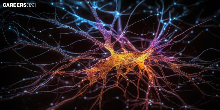
A neuron is a kind of cell in the nervous system that transmits signals for communication within the body. It is important to many functions, such as sensation, movement, and reflexes. This is one of the important topics in Class 11 under the chapter Neural Control and Coordination from which questions are asked in competitive exams like NEET and AIIMS BSc Nursing where biology is one of the main subjects.
This Story also Contains
- Neuron Definition
- What is a Neuron?
- Structure of a Neuron
- Types of Neurons
- Functions of Neurons
- Recommended Video on Neuron
Neuron Definition
A neuron is a specific cell of the nervous system that carries electrical impulses along the pathway of the nerve, by which the brain, spinal cord, and other parts of the body can communicate with one another.
What is a Neuron?
The neuron is the main part of the brain and the nervous system. They participate in the reception, processing and transmission of sensory information and motor commands to the muscles, thus they control various body functions. These are the parts of nature that have specialised cells that transfer electrical and chemical signals. Hence, they are very much responsible for the nervous system's proper functioning.
Also Read:
- Neural Control and Coordination
- Central Nervous System
- Peripheral Nervous System
- Chemical Coordination and Integration
Commonly Asked Questions
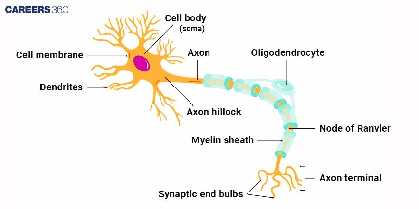
Structure of a Neuron
Knowing the general structure of a neuron will help us appreciate how it works in the nervous system.
Cell Body (Soma): The portion that includes the nucleus, regarded as the control centre of the cell.
Dendrites: These are incoming signal-receiving ends from other neurons.
Axon: Conducts electrical impulses away from the cell body
Myelin Sheath: Fatty, insulating layer of the axon, increasing the velocity of signal transmission.
Nodes of Ranvier: These are gaps in the myelin sheath that facilitate rapid transmission of signals.
Axon Terminals: These are responsible for releasing neurotransmitters to pass on the signals to the next neuron.
Diagram: Structure Of Neuron
Commonly Asked Questions
Types of Neurons
There can be three primary types of neurons based on their functions.
Sensory Neurons: These carry sensory information to the brain and spinal cord.
Motor Neurons: These transmit commands from the brain and spinal cord to muscles and glands.
Interneurons: These are neurons that connect other neurons, usually within the central nervous system. This means they connect sensory with motor neurons, therefore, facilitating communication between them.
Commonly Asked Questions
Functions of Neurons
The neurons perform various crucial functions which help the nervous system function properly.
Signal Transmission: The neurons transmit electrical impulses called action potential.
Synaptic Transmission: It is the process through which neurotransmitters are released to communicate with other neurons.
Neurotransmitters
Thus, neurotransmitters are the chemical mediators of the signals transmitted from one neuron to another.
Dopamine: Participates in mechanisms related to reward and pleasure.
Serotonin: Modulates mood and social behaviour.
Acetylcholine: Involved in the action of muscles and memory.
The Synapse
The synapses are the junctions through which neurons communicate with each other.
The presynaptic terminal, synaptic cleft, postsynaptic membrane.
It allows the transmission of electrical or chemical signals from one neuron to another.
Neurogenesis and Neuron Plasticity
It is in charge of learning and recovery; it relies on the generation of new neurons and plasticity in the brain.
Neurogenesis: The process of new neuron generation in the Brain.
Neuron Plasticity: Changes a neuron undergoes as a result of new information, sensory stimulation, development, damage, or malfunction.
Neuron-related Disorders
Several neurological diseases are claimed to be connected to the failure of neurons.
Alzheimer’s Disease: A condition characterised by the death of neurons, and the consequences are memory loss and cognitive declines
Parkinson’s Disease: Involves the loss of dopamine-producing neurons, affecting movement and coordination.
Multiple Sclerosis: An autoimmune disorder that attacks the myelin sheath, disrupting neural communication.
Also Read:
| Autonomic Nervous System | Sympathetic Nervous System |
| Difference Between Brain and Spinal Cord | Reflex Action and Reflex Arc |
| Nerve Fibres | Cerebrospinal Fluid |
Commonly Asked Questions
Recommended Video on Neuron
Frequently Asked Questions (FAQs)