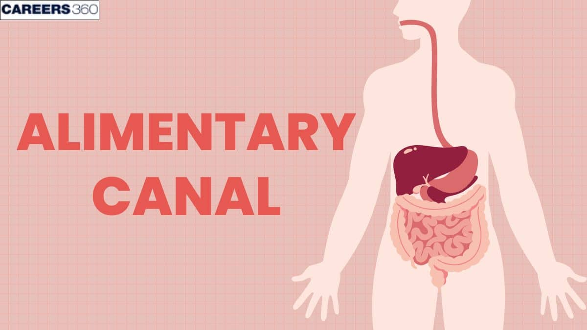
Alimentary canal - Example, Defination, Diagram
The alimentary canal is the long digestive tube in humans and animals, enabling food breakdown, nutrient absorption, and waste elimination. Learn its parts, functions, disorders, and variations in different species.
This Story also Contains
- Introduction to the Alimentary Canal
- Segments of the Human Alimentary Canal
- Main Parts of the Alimentary Canal
- Primary and Supplementary Functions
- Common Disorders of the Alimentary Canal
- Comparative Anatomy - Alimentary Canals in Different Animals
- Tips, Tricks, and Mnemonics for Learning
- Exam Relevance and Weightage
- Alimentary Canal NEET MCQs
- FAQs on Alimentary Canal
- Recommended Video on Alimentary Canal

Introduction to the Alimentary Canal
The food we eat contains a variety of nutrients, which are used for building new body tissues and repairing damaged tissues. Food is also vital to life because it is our only source of chemical energy. However, most of the food we eat consists of molecules that are too large to be used by body cells. Therefore, foods must be broken down into molecules that are small enough to enter body cells for their use. This is accomplished by the digestive system. The combination of extensive environmental exposure and close association with blood vessels is essential for processing the food that we eat.
Segments of the Human Alimentary Canal
The alimentary canal, or digestive tract, is a long tube that forms a continuous pathway through which food passes as it undergoes digestion and absorption. The entire length of the canal measures approximately 9 meters in length, from the mouth to the anus. It begins in the mouth by mechanical and chemical breakdown, and ends with the absorption of nutrients in the small intestine, besides elimination from the anus.
The primary segments of the human alimentary canal are:
Segment | Length | Function |
15 cm | Mechanical digestion (chewing), salivary secretion | |
25 cm | Transportation of food to the stomach | |
Stomach | 25 cm | Chemical digestion, mixing of food with gastric juices |
6 meters | Nutrient absorption | |
1.5 meters | Water absorption, faeces formation | |
Rectum | 15 cm | Storage of faeces |
Anus | 3 cm | Excretion of waste |
Main Parts of the Alimentary Canal
The three main parts of the human alimentary canal are
Oral cavity
Transport passage
The digestive tract
Oral cavity
The oral cavity is where food is swallowed. It is made up of tongue, teeth, and palate.
Additionally, the teeth's movement breaks down the food into tiny pieces.
Human saliva in the mouth cavity softens the food. Amylase and lysozyme found in saliva are released by the salivary gland.
Food is moved more easily across the oral cavity thanks to the motion of the jaws and tongue.
The foodstuff bolus is moistened and broken up after which it is delivered to the oesophagus via deglutition (swallowing).
Transport Cavity (Esophagus)
The transport channels do not cause any biochemical changes in the food bolus, they merely make it easier for it to move via peristalsis from one section of an alimentary canal to the next.
These transport passageways are essentially muscular tubes with stratified squamous epithelium and a few mucous glands that produce lubricating mucus lining the inside.
Digestive Tract
The digestive system is further divided into many parts
Stomach
Small intestine
Large intestine
Primary and Supplementary Functions
Ingesting food material and separating it into smaller portions is the main purpose of the alimentary canal or digestive tract. The tiny fractions that are transformed into smaller molecules by a number of secretions, primarily digestive enzymes, are taken up by the blood and lymphatic systems.These small molecules, which serve as the building blocks for the creation of vital proteins, carbohydrates, and lipids, primarily consist of amino acids, short peptides, sugars, and fatty acids.
The digestion of food and the absorption of nutrients from it are the main tasks of the alimentary canal. The alimentary canal also serves various secondary but crucial duties in addition to these basic ones. These are what they are:
For a variety of dangerous bacteria, the alimentary canal serves as an immunological barrier. Gut-associated lymphoid tissue (GALT) and the various pH conditions that exist throughout the alimentary canal perform this role.
Additionally, intestinal bacteria support immunological homeostasis.
The colonic bacterial colony also hinders the development of dangerous germs in our gastrointestinal tract.
Additionally, drug metabolism takes place in the alimentary canal, where the drug molecule is broken down into smaller pieces before being expelled from the body.
Common Disorders of the Alimentary Canal
Several disorders may remodel the functioning of the alimentary canal and lead to various symptoms and health issues.
Gastroesophageal Reflux Disease (GERD)
Symptoms: Acid reflux, heartburn, regurgitation
Cause: Malfunction of the lower oesophagal sphincter that normally prevents the backward flow of stomach acid from the stomach into the oesophagus.
Peptic Ulcers
Symptoms: Abdominal pain, bloating, nausea.
Cause: Chipped lining of stomach or duodenum usually after infection with H. pylori, or with prolonged NSAIDs.
Irritable Bowel Syndrome
Symptoms: abdominal pain, cramping, bloating, changes in bowel habits.
Cause: it includes altered gut-brain interaction, disorders of gut motility, and enhanced sensitivity.
Crohn's Disease
Symptoms: Chronic inflammation, diarrhoea, abdominal pain, and weight loss
Cause: This is an autoimmune disorder that makes the body attack any part of the gastrointestinal tract. Therefore, it causes chronic inflammation and damage.
Comparative Anatomy - Alimentary Canals in Different Animals
The alimentary canal of different animals has a wide range of anatomical differences. While some animals only have a single chamber in their stomach, others have many chambers (multiple-chamber stomachs).
Rabbits
Rabbits have larger caecum and small intestines to provide a broad surface area again for nutrient absorption from plant matter.
Birds
Birds have a gizzard, a proventriculus, and a two-chambered stomach. The proventriculus employs stomach fluids to aid in digestion, as opposed to the gizzard where food is mechanically broken down, stored, and then later digested again. Furthermore, birds vomit up uneaten food.
Due to the absence of fangs in their mouth cavity, birds do not masticate their food. Given that their beaks are made to be used for chewing food, several bird species have unique beak shapes. While waste is ejected through the cloaca or entrance, nutrients are absorbed in the small intestine.
Ruminants
Ruminants with several stomachs include sheep, cattle, goats, and others. The primary goal of this adaptation is to help ruminants digest dietary cellulose, which makes up the majority of their diet of plants. There are four compartments that make up the stomach which are as follows :
Rumen
Reticulum
Omasum
Abomasum
The abomasum is viewed as being equivalent to a mono-gastric stomach or entire stomach because it secretes gastric juices.
Similar to how ruminants and reticulum break down cellulose in food, the stomach is filled with microbial flora that aids in digestion. Additionally, the food that is consumed undergoes fermentation in these chambers, creating a substantial amount of fumes.
These glasses are occasionally released by the animal. Ruminants rechew and regurgitate the food substance in the reticulum chamber before moving it to the omasum, the fourth chamber.
The omasum expels water. After that, the food is sent from the omasum to the abomasum for enzyme and gastric secretion digestion before being eventually transferred to the small bowel. Nutrients are taken up in the small intestine, and waste is removed from the body through the large intestine.
Pseudo-Ruminants
Pseudo-ruminants, like camels, have multiple stomachs in contrast to ruminants. Camels don't have a rumen. The camel's stomach has three compartments: the omasum, abomasum, and reticulum. It's interesting to note that camels have larger Caecum. Camels have a Caecum which is rich with microbial flora and is in charge of breaking down the plant-based roughage that makes up the majority of their diet.
Cockroach
The alimentary canal of cockroaches divides into three sections and extends from the mouth to the anus. The pharynx, oesophagus, crop, and gizzard make up the anterior stomodaeum (foregut). Middle stomach Proctodaeum in the posterior hindgut The intestine (or small bowel), stomach (or intestine), and rectum make up this.
Earthworm
The alimentary canal of an earthworm is a straight tube with specialized regions:
Pharynx: For swallowing.
Esophagus: For transport.
Crop: For storage.
Gizzard: For grinding food.
Intestine: For absorption.
Tips, Tricks, and Mnemonics for Learning
Study aids: Use diagrams or flowcharts to visualize each part's structure and functions.
Use Mnemonics. You may find it easier to remember the structures if you recall: M.E.S.S.I.L.R.A, Mouth, Esophagus, Stomach, Small Intestine, Large Intestine, Rectum, Anus.
Multimedia Learning: Watch videos or animations showing the process of digestion to get a better grasp.
Analogies Using: An example to remember is the alimentary canal compared to a food processing factory.
Flashcards: These are a great way to make flashcards for keywords like peristalsis, bile, or absorption and quickly revise them.
Link to Real Life: Relate the alimentary canal to common health conditions, such as indigestion or acid reflux, to be able to understand it better in real-life applications.
Exam Relevance and Weightage
Here's a summary of the weightage of questions related to the alimentary canal in various exams:
Alimentary Canal NEET MCQs
Q1. Which of the following is not a digestive gland?
Salivary glands
Pancreas
Liver
Gallbladder
Correct answer: 4) Gallbladder
Explanation:
The GI System is concerned with the processes of digestion and absorption of nutrient substances essential to life. This system includes an alimentary canal, which represents a hollow organ beginning at the mouth and the oesophagus, stomach, and small intestine divided into the duodenum, jejunum, and ileum, the large intestine representing the colon and rectum culminating in the anus. Glands associated with the digestive glands include the salivary gland, pancreas, and liver, but the gall bladder stores bile to be used within the digestive pathway.
Hence the correct answer is Option (4) Gallbladder.
Q2. The GI tract or alimentary canal in humans starts with the ________ and proceeds to the __________, stomach, small intestine, and then to the large intestine (colon), rectum, and terminates at the _________. Fill in the blanks by choosing the correct option.
Tongue, trachea, anus respectively
Mouth, pharynx, duodenum respectively
Mouth, esophagus, anus respectively
Salivary glands, larynx, jejunum respectively
Correct answer: 3) Mouth, esophagus, anus respectively
Explanation:
The gastrointestinal (GI) tract, also known as the alimentary canal starts with the mouth and proceeds to the esophagus, stomach, small intestine (duodenum, jejunum, ileum), and then to the large intestine (colon), rectum, and terminates at the anus. It has an endodermal origin during embryonic development.
Hence, the correct answer is option 3) Mouth, oesophagus, and anus respectively.
Q3. Lamina propria is
Layer of loose connective tissue
It is a part of Mucosa layer of Alimentary canal
Also contains gland for the secretion of gonadotropin hormones
Both 1 and 2
Correct answer: 4) Both 1 and 2
Explanation:
Mucosa: It is composed of simple epithelium cells and a thin connective tissue - areolar tissue. The layer of areolar connective tissue is called lamina propria. In addition, the mucosa has a thin, smooth muscle layer, called the muscularis mucosa. Mucosa forms glands in the stomach (gastric glands) and crypts in between the bases of villi in the intestine (crypts of Lieberkuhn).
Hence, the correct answer is option 4) Both 1 and 2.
Also Read:
FAQs on Alimentary Canal
Q1. What is the alimentary canal and what are its main functions?
The alimentary canal is a long, muscular tube of 8-9 meters long that starts from the mouth and ends at the anus. It is the main part of the digestive system. The major functions of the digestive system are:
1. Ingestion: taking food into mouth.
2. Secretion: release of water, acid, buffers, and enzymes into lumen of GI tract.
3. Mixing and propulsion: churning and movement of food through GI tract.
4. Digestion: mechanical and chemical breakdown of food.
5. Absorption: passage of digested products from GI tract into blood and lymph.
6. Defecation: elimination of feces from GI tract.
Q2. What are the primary segments of the human alimentary canal?
The primary segments of the human alimentary canal are:
Mouth - for ingestion and chewing of the food
Pharynx - acts as a passage between moth and oesophagus.
Oesophagus - muscular tube that carries food to stomach via peristalsis movement
Stomach - digest food, especially proteins
Small intestine - main site of digestion and nutrient absorption
Large intestine - absorbs water and salts
Anus - ejects the waste material from the body.
Q3. How does food move through the transport passage of the alimentary canal?
Food enters the mouth and then is moved in the esophagus by peristalsis movement. It is a series of contractions and relaxations of smooth muscles in the canal walls. It begins at the middle of the oesophagus and continues throughout the alimentary canal. It makes sure that the food is pushed forward, mixed with the digestive juices and the enzymes properly. The food sometimes flows backwards and sphincters are present to prevent this backward flow.
Q4. What are common disorders of the alimentary canal?
Some of the common disorders of the alimentary canal are:
Indigestion – discomfort due to incomplete digestion.
Constipation – difficulty in passing stools due to hard feces.
Diarrhea – frequent watery stools caused by infection or food intolerance.
Peptic ulcers – sores in stomach or duodenum lining due to excess acid or bacterial infection.
Irritable Bowel Syndrome (IBS) – functional disorder causing abdominal pain, bloating, and irregular bowel habits.
Recommended Video on Alimentary Canal
Frequently Asked Questions (FAQs)
Goblet cells are modified epithelial cells that secrete mucus, aiding in lubrication and protection of the intestinal lining.
The movement is caused by peristalsis, which involves the contraction of smooth muscles lining the canal.
The bile enters the duodenum, the first part of the small intestine.
The primary function of the alimentary canal is to break down food into absorbable components and transfer those components to the different organs of the body
The organs through which substances are transported before being consumed, broken down, absorbed, and discharged as feces. These include the rectum, anus, pharynx (throat), esophagus, gut, small bowel, digestive tract, and lips.