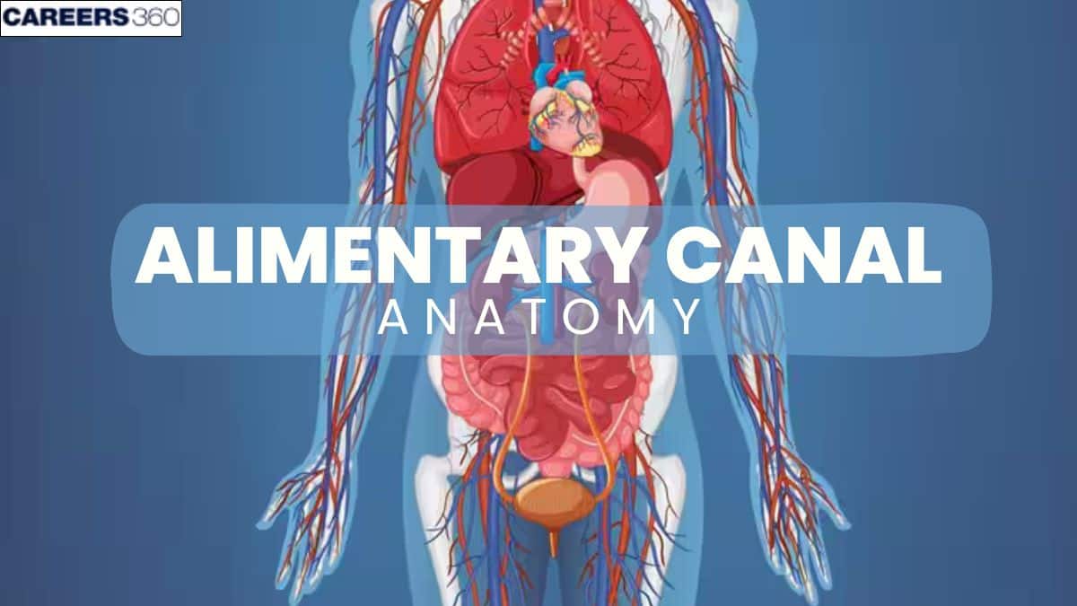
Alimentary Canal - Anatomy, Structure, Functions, Types
The alimentary canal is often referred to as the digestive tract. It is long, tube-like with no apparent lumen. It processes food through mechanical and chemical digestion, absorbs nutrients, and eliminates waste.Understanding its parts and layers is essential for NEET, Class 11 and 12 Biology, and medical entrance exams.
This Story also Contains
- Introduction to the Alimentary Canal
- Parts of the Alimentary Canal
- Parts of the Alimentary Canal
- Layers of the Alimentary Canal
- Alimentary Canal NEET MCQs
Introduction to the Alimentary Canal
The alimentary canal is an extended, continuous, hollow tube running from the mouth to the anus. It performs the digestion of food and absorption of nutrients, with the elimination of waste. The complex system involves multiple organs and processes working to ensure that the body receives proper nourishment for energy, growth, and repair. The anatomy and functions of the alimentary canal are the keys to understanding how our bodies process the food we eat.
Parts of the Alimentary Canal
The alimentary canal is about a 9-meter-long, continuous muscular tube extending from mouth to anus. It represents an important part of the digestive process, consisting of many organs that are specially differentiated regions of this canal, working in a coordinated manner on food digestion, absorbing the released nutrients, and finally, the elimination of the remaining waste.
Those sections are the mouth, pharynx, oesophagus, stomach, small intestine, large intestine, rectum, and anus. All these sections take part in digestion and nutrient absorption to meet the organism's needs for proper functioning. An adult alimentary canal is approximately 9 meters in length.
Alimentary Canal
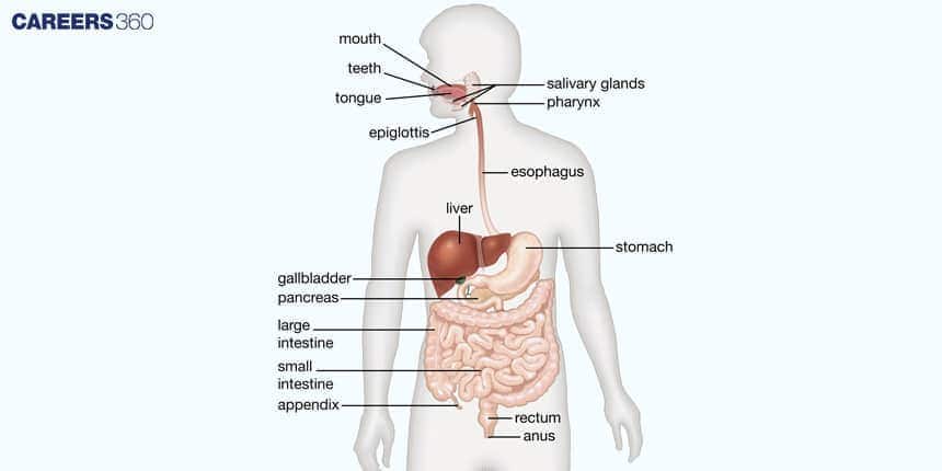
Parts of the Alimentary Canal
Each part of the alimentary canal has different structures and carries out different functions required in digestion.
Mouth
Teeth: Aids in mechanical digestion by grinding.
Tongue: Aids in moving food around with its movement and is covered with taste buds.
Salivary Glands: Secrete saliva that contains an enzyme amylase.
Function
Ingestion: Taking food.
Mastication: The mechanical breaking of food by chewing.
Enzymatic Breakdown: Initial digestion of carbohydrates by salivary amylase.
Pharynx
Oropharynx: The passageway for food.
Nasopharynx: Part of the respiratory pathway.
Laryngopharynx: It leads to the oesophagus.
Function
Pathway for Food and Air: The food passes into the oesophagus and the air into the larynx.
Oesophagus
The lining of the muscular tube is of stratified, squamous epithelium.
The Sphincters: There is an upper oesophagal sphincter and a lower oesophagal sphincter.
Function
Peristalsis: Wave contractions of the muscles which propel the food towards the stomach.
Transport: Food from the pharynx into the stomach.
Stomach
Regions: Fundus, body, antrum, and pylorus
Gastric Glands: Secrete hydrochloric acid and pepsinogen
Function
Storage: Stores swallowed food temporarily.
Mixing: Food is churned with gastric juices and changed into chyme.
Acid Secretion: Hydrochloric acid activates the pepsinogen into pepsin.
Initial Protein Digestion: Pepsin breaks down protein into peptides.
Small Intestine
Segments: Duodenum, jejunum, ileum.
Villi and Microvilli: Increase surface area to absorb.
Function
Absorption of Nutrients: Primary area for the absorption of nutrients into the bloodstream.
Digestion: Further break down the carbohydrates, proteins, and fats.
Surface area enhancement: The villi and microvilli maximise the efficiency of absorption.
Large Intestine
Segments: Cecum, colon , rectum.
Houses some friendly bacteria.
Function
Water Absorption: Indigestible food has water and electrolytes reabsorbed here.
Formation of Feces: The residual waste is compacted into faeces.
Vitamin Production: Some vitamins, such as vitamin K, are produced by bacteria.
Rectum and Anus
Rectal Walls: There are stretch receptors that send signals that it is time to defecate.
Sphincters in Anus: There are internal ( involuntary ) and external ( voluntary ) sphincters.
Function
Storage: This is the final storage area for faeces until it is eliminated.
Defecation: Controls the expulsion of faeces from the body.
Layers of the Alimentary Canal
The alimentary tract's walls share a fundamentally consistent basic structure across the whole tract. To achieve the necessary functionality, specific portions of an alimentary canal may, however, differ physically. The human alimentary canal tract is further divided into four layers from inside to outside respectively :
Mucosa (Innermost layer)
Sub Mucosa
Muscularis
Serosa (Outermost layer)
Mucosa
The mucosal, or mucous membrane layer, is the innermost wall lining the alimentary canal lumen. Three more layers are separated from the mucous membrane Epithelium, Lamina propria which is a loose connective tissue layer, and Muscularis mucosa- a smooth muscle layer.
Different portions of the alimentary canal have differences in their fundamental structure depending on their activity and role.
The mucosal membrane in the mouth and the anus is composed of stratified squamous tissue to protect the alimentary canal from abrasion, while the stomach and intestine have thin columnar cells in the epithelium to allow for nutrient absorption.
The epithelium of the small intestine is folded into finger-like extensions to create a high surface area for nutrient absorption.
In the mucus layer, epithelial tissue like goblet cells release mucus and specific digestive enzymes.
Submucosa
The mucosal membrane is encircled by an uneven, thick layer of connective tissue that houses lymphatic vessels, nerve terminals, and blood vessels.
The submucosal layer is made up of fibroblasts, collagen and an acellular matrix. This layer also contains certain glands, such as Brunner glands.
The submucosa of the duodenum is the site of Brunner's glands.
They release an alkaline fluid that contains mucin that protects the mucosa from the stomach contents' acidic influx into the duodenum.
Muscularis is a layer of smooth muscle that is in charge of the peristaltic movement and segmental contractions that carry food along the alimentary canal.
Its longitudinal muscle layer as well as the inner circular layer are the two layers in which these smooth muscles are organized.
Between the two smooth muscle layers is a small layer of connective tissue.
The plexus Myenteric plexus or also called as Aurbach plexus.
It is a type of vascular connective tissue that houses the myenteric nerve plexus.
The motility of the digestive tract is controlled by the myenteric nerve plexus.
Muscularis
The muscularis of the mouth, pharynx, and superior and middle parts of the esophagus contains skeletal muscle that produces voluntary swallowing.
Skeletal muscle also forms the external sphincter of anus, which permits voluntary control of defecation.
The rest of the tract, the muscularis, consists of smooth muscle that is generally found in two sheets, an inner sheet of circular fibers and an outer sheet of longitudinal fibers.
Involuntary contractions of the smooth muscle help break down food, mix it with digestive secretions, and propel it along the tract. Between the layers of the muscularis is a second plexus of neurons—the myenteric plexus
Serosa
The organs located in the peritoneal cavity behind the diaphragm are covered by the serosa, a loose layer of connective tissue.
This layer, which is made up of secretory epithelial cells, lessens friction brought on by muscle activity.
Alimentary Canal NEET MCQs
Q1. The alimentary canal is composed of four layers of tissues. Out of these four, the one containing the nerve plexus is
Mucosa
Submucosa
Serosa
Muscularis
Correct answer: 2) Submucosa
Explanation:
There are four levels in the alimentary canal. The layer that houses the nerve plexus is called the submucosa. There are lymphatics and blood arteries in this stratum as well. The submucosa's nerve plexus aids in regulating processes including secretion and digesting. Each of the other layers—the mucosa, muscularis externa, and serosa—contributes to the movement and digestion of food.
Hence, the correct answer is option 2) Submucosa.
Q2. Starting from the outer side to the inner, the correct sequence of layers of the alimentary canal is represented as
Muscularis, submucosa, mucosa, and serosa
Submucosa, muscularis, mucosa, and serosa
Serosa, muscularis, submucosa, and mucosa
Mucosa, submucosa, muscularis, and serosa
Correct answer: 4) Mucosa, submucosa, muscularis, and serosa
Explanation:
The correct order of layers in the alimentary canal, from outside inwards, is:
1. Serosa/Adventitia: The exterior layer, a connective tissue membrane, shields digestive organs and offers structural support.
2. Muscularis externa: Composed of two smooth muscle sub-layers (circular and longitudinal), it aids in peristalsis, moving food along the tract.
3. Submucosa: Beneath this muscular layer is connective tissue with blood, lymph, and nerve vessels, supporting the mucosa and supplying nutrients.
4. Mucosa: The inner lining directly interacting with digestive content includes:
a. Epithelium with goblet cells secreting mucus for lubrication.
b. Lamina propria, a thin connective tissue for support.
c. Muscularis mucosa, a fine layer of smooth muscle.
Hence, the correct answer is option 4) Mucosa, submucosa, muscularis, and serosa.
Q3. Which is true regarding Auerbach Plexus?
It is also known as Meissner Plexus
Lies in Muscularis layer
Lies in submucosal layer
Responsible for regulating secretions
Correct answer: 2) Lies in Muscularis layer
Explanation:
Auerbach’s plexus, also called the myenteric plexus, is located between the circular and longitudinal muscle layers of the muscularis externa in the gastrointestinal tract. It is responsible for controlling peristalsis and the motility of the gut. It is different from Meissner’s plexus, which lies in the submucosal layer and regulates glandular secretions and local blood flow. Confusing the two is common, but their locations and functions are distinct — Auerbach’s for movement, Meissner’s for secretion control.
Hence, the correct answer is option 2) Lies in the Muscularis layer.
Also Read:
FAQs on Alimentary Canal
Which organs make up the human alimentary canal?
The human alimentary canal is 8-9 meters long, starting from mouth to the anus. It includes the following organs:
Mouth - for ingestion and chewing of the food
Pharynx - acts as a passage between moth and oesophagus.
Oesophagus - muscular tube that carries food to stomach via peristalsis movement
Stomach - digest food, especially proteins
Small intestine - main site of digestion and nutrient absorption
Large intestine - absorbs water and salts
Anus - ejects the waste material from the body.
What role do teeth, tongue, and salivary glands play in digestion?
Teeth - Humans have 32 permanent teeth. They are divided into four types based on the function they perform i.e., incisors, canines, premolars, and molars. They perform mastication, break food into smaller pieces for easy digestion and swallowing. They are also adapted for cutting, tearing, and grinding food.
What are the main regions of the stomach and their functions?
The stomach has four main regions:
Cardiac region – near the oesophagus, receives swallowed food.
Fundus – dome-shaped upper part; stores food temporarily.
Body – main central region; mixes food with gastric juices.
Pyloric region – lower part; regulates passage of chyme into the duodenum through the pyloric sphincter.
Functions of the stomach:
Secretes gastric juice containing HCl, pepsinogen, and mucus.
Performs mechanical churning and mixing of food.
Begins protein digestion using pepsin.
Acts as a temporary food reservoir.
What are the four main layers of the alimentary canal wall?
The four main layers of the alimentary cana from innermost to outermost are -
Mucosa – inner lining which secretes mucus, digestive enzymes, and hormones, contains folds and villi for absorption.
Submucosa – connective tissue layer with blood vessels, lymph vessels, and nerves.
Muscularis – smooth muscle layer (inner circular and outer longitudinal layers) responsible for peristalsis.
Serosa – outermost protective layer made of thin epithelium and connective tissue, forms part of the visceral peritoneum.
Recommended Video on Alimentary Canal
Frequently Asked Questions (FAQs)
The small intestine has villi and microvilli that increase the surface area for efficient digestion and absorption of nutrients.