Areolar Connective Tissues
Areolar connective tissue is a loose connective tissue found beneath the skin, around organs, muscles, blood vessels, and nerves. It provides strength, elasticity, nutrient storage, and immune defense. Class 11 Biology and NEET focus on its structure, cell types, fibers, and clinical relevance.
This Story also Contains
- What is Areolar Connective Tissue?
- Structure and Composition of Areolar Connective Tissue
- Location of Areolar Connective Tissue in the Body
- Functions of Areolar Connective Tissue
- Disorders and Diseases Of Areolar Connective Tissue
- Areolar Connective Tissue NEET MCQs (With Answers & Explanations)
- Recommended Video On 'Areolar Connective Tissue'
What is Areolar Connective Tissue?
Areolar connective tissue is one of the most widely distributed connective tissues. It is a loose connective tissue present under epithelia and between other tissues. It consists of fibers arranged randomly and several kinds of cells like fibroblasts, macrophages and a few white blood cells, embedded in semifluid ground substance. Its functions as a universal packing material between other tissues, providing support and elasticity, and playing a vital role in the immune response and nutrient storage.
Structure and Composition of Areolar Connective Tissue
Areolar connective tissue is a form of connective tissue that consists of a variety of cells, fibres, and amorphous ground substances. This tissue is somewhat flexible and supportive.
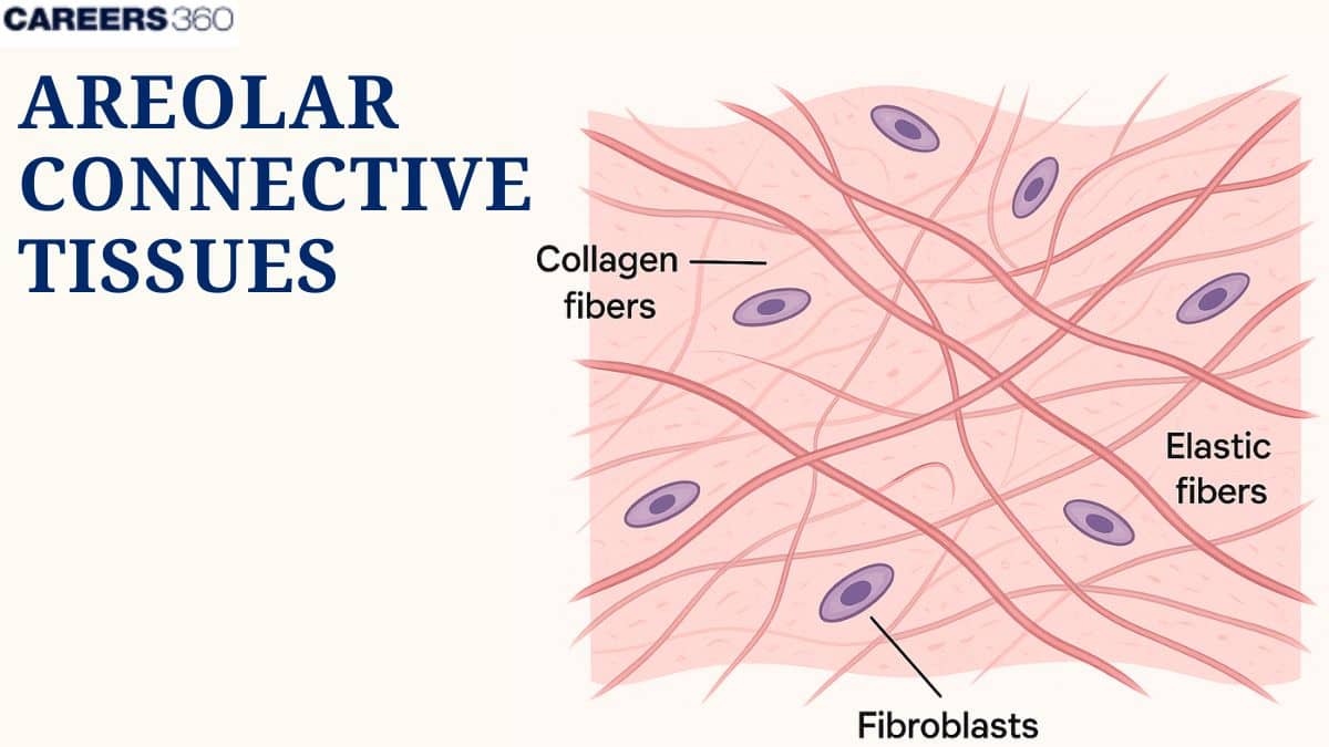
Cell Types
The most common cells in areolar connective tissue are fibroblasts, macrophages, plasma cells, adipocytes, mast cells and white blood cells.
Fibroblasts
It produces extracellular matrix and collagen. It provides structural framework and helps in the healing of wounds.
Macrophages
These phagocytose and digest pathogens and debris. They participate in immune defence and produce cytokines to modulate immune responses.
Mast Cells
They contain histamine and heparin-rich granules. These play a role in allergic reactions and are also involved in wound healing and defence against pathogens.
White Blood Cells
WBCs include lymphocytes, neutrophils, and eosinophils. They contribute to immune response and help in fighting infections.
Fibres
The fibres in the areolar connective tissue provide strength, elasticity, and support.
Fibres | Explanation |
Collagen Fibers |
|
Elastic Fibers |
|
Reticular Fibers |
|
Ground Substance
Ground substance is a gel-like substance that fills the space between many cells and fibers. It contains proteoglycans, glycosaminoglycans, and glycoproteins. It is also rich in water and electrolytes. The functions iclude:
Provides a medium for the exchange of nutrients and waste.
Lubricates and protects cells.
Allows cell migration.
Location of Areolar Connective Tissue in the Body
Areolar connective tissue is distributed in the body at places where support and flexibility are required.
Subcutaneous Layer
Underlying skin
Connects skin to underlying tissue.
Provides insulation and cushioning.
Around Organs
Surrounds internal organs.
Provides support and holds organs in place.
Allows for flexibility and movement.
Between Muscles
Fills spaces between muscle fibers.
Supports muscle function.
Permits muscle movement without friction.
Around Blood Vessels And Nerves
Surrounds blood vessels and nerves.
Provides protection and support.
Ensures flexibility and movement.
Functions of Areolar Connective Tissue
Areolar connective tissue performs several vital functions in the body, including binding and support, nutrient storage, and immune defence.
Binding and Support
Areolar connective tissue performs the function of connecting and supporting different tissues and organs.
Holding skin to underlying tissues
The skin is attached to muscles and bones.
Gives support and flexibility.
The internal organs are surrounded and cushioned.
Organ position is maintained, and at the same time, movement is allowed.
Nutrient Storage
Areolar connective tissue stores nutrients, mainly in the form of fat cells.
They store energy in the form of lipids.
They provide insulation and cushioning.
Upon requirement, they release energy.
Immune Defense
Areolar connective tissue helps to provide the body's immune response.
Initiates inflammation to guard against injury and infection.
Histamine release by mast cells.
Phagocytosis by macrophages
Macrophages ingest and digest pathogens and debris.
Aids in the cleaning of infection sites and healing.
Development and Repair of Areolar Connective Tissue
Tissue development and repair processes involve areolar connective tissue.
Embryonic Development
Origin from Mesenchymal Cells
Differentiation of mesenchymal cells results in areolar connective tissue.
Form the basis of connective tissues in the body.
Wound Healing and Tissue Regeneration
Role In Tissue Repair And Regeneration
Fibroblasts produce collagen and extracellular matrix.
Promote tissue regeneration and repair.
Stages of Wound Healing Involving Areolar Tissue
Inflammation: Initial response to injury
Proliferation: Formation of new tissues
Remodeling: Maturation and strengthening of new tissue
Disorders and Diseases Of Areolar Connective Tissue
Areolar connective tissue can be affected by various disorders and diseases
Inflammatory Conditions
It occurs when pathogens trigger immune cells in the tissue i.e., overactive inflammatory response
This causes swelling, redness and pain in the area. Involve overactive inflammatory response
Examples include local infections, allergy, asthma
Fibrosis
It is the abnormal or excessive proliferation of fibrous connective tissue
It replaces normal areolar tissue with scar tissue which has reduced flexibility.
It is common in organs like lungs, liver and skin and may lead to organ failure.
Areolar Connective Tissue NEET MCQs (With Answers & Explanations)
Important topics for NEET exams:
Types of Cell and their Functions
Types of Fibres (Collagen, Elastic, Reticular)
Location of Areolar Tissue
Practice Questions for NEET
Q1. Large amoeboid cells that are a part of our intimate immune system, found in areolar tissue are called as
Macrophytes
Mast Cells
Fiboblasts
Adipophytes
Correct answer: 1) Microphytes
Explanation:
Areolar tissue is a type of loose connective tissue found beneath the skin and between organs. It provides support, elasticity, and strength while allowing movement. The cells present in areolar tissue are Macrophages, fibroblasts, and mast cells.
Hence, the correct answer is option 1) Macrophytes
Q2. Ligaments are a type of _____ tissue.
Areolar Connective tissue
Adipose Connective tissue
Skeletal Connective tissue
Fluid
Correct answer: 1) Areolar Connective tissue
Explanation:
The ligament is not a type of areolar connective tissue. The ligaments consist of dense regular connective tissue, which is tightly packed in parallel collagen fibres, giving them strength and stability to the joint by connecting bones to other bones. They do not resemble the areolar connective tissue that is loose and flexible, meant to support and bind other tissues. Ligaments are essential in stabilizing the joints and restricting excessive movement.
Hence, the correct answer is option 1) Areolar Connective tissue.
Q3. Choose the incorrect statement
Areolar connective tissue is the most widely distributed connective tissue.
Areolar connective tissue contains cells such as fibroblasts, mast cells, macrophages, lymphocytes, plasma cells, mesenchyme cells, chromatophores, fat cells.
Areolar connective tissue acts as a fat reserve
Areolar connective tissue can allow the diffusion of materials and cells to the infected area
Correct answer: 3) Areolar connective tissue acts as a fat reserve
Explanation: Functions:
The major function of areolar connective tissue is to bind the parts together.
It also provides strength, elasticity, and support.
It can also allow the diffusion of materials and cells to the infected area.
Areolar connective tissue does not act as fat reserve.
Hence, the correct answer is option 3) Areolar connective tissue acts as a fat reserve.
Recommended Video On 'Areolar Connective Tissue'
Frequently Asked Questions (FAQs)
Areolar connective tissue plays a key role in inflammation by housing mast cells that release histamine, initiating the inflammatory response, and macrophages that engulf pathogens and debris.
The normal functions of areolar connective tissue include supporting and providing elasticity to other tissues, acting as a packing material, storing nutrients, and contributing to immune defences.
In the body, the areolar connective tissue is located in the dermis of the skin, surrounding organs, in the space between muscles, and around blood vessels and nerves.
Areolar connective tissue contains fibroblasts, macrophages, mast cells and white blood cells.
Areolar connective tissue is loose and has a more open structure with fewer fibres than dense connective tissue that has tightly packed collagen fibres, therefore providing greater strength but less flexibility.