Animal Tissue - Definition, Characteristics, Types, Question Types, Tips
Animal tissue refers to collections of specialized cells organized for specific functions. There are four major types – epithelial, connective, muscular, and nervous, each with unique roles in protection, movement, support, and coordination. Learn their structure, functions, differences, and NEET exam questions.
This Story also Contains
- What Is Animal Tissue?
- Characteristics of Animal Tissue
- Types of Animal Tissue
- Epithelial tissue
- Muscular tissue
- Connective Tissue
- Nervous tissue
- Difference Between Plant Tissue and Animal Tissue
- Animal Tissue Culture Techniques
- Tips, Tricks, & Strategies to Study Animal Tissue
- Exam Weightage & Types of Questions
- Animal Tissue NEET MCQs
- FAQs on Animal Tissue
Animal tissue is a term referring to specialized cells designed to do a key job to keep living. During embryonic development, the germ layers become differentiated into four kinds of tissues. These are epithelial, connective, muscular, and nervous tissues. Animal tissues help to understand the structural organisations in animals. Knowing the difference between plant and animal tissue is important because tissues involving animals do not have cell walls and are more complex. Also, knowledge about animal tissue culture techniques is also very significant in research and for their medical applications.
What Is Animal Tissue?
Animal tissue refers to any collection of specialized cells organized together for specific functions within an organism. Such tissues are critical for general construction and functioning in animals because they provide the building blocks of organs and systems. The four major categories of animal tissues include epithelial, connective, muscular, and nervous tissues. Each type has peculiar characteristics and functions, for example, epithelial tissue protects and covers surfaces, connective tissue supports and binds other tissues, muscular tissue facilitates movement and nervous tissue transmits signals throughout the body. Knowing what animal tissue is and the several forms it can take constitutes the essence of understanding animal biology and physiology. Understanding what animal tissue is helps in comprehending how different tissues combine to form organs and organ systems. The study of what animal tissue is crucial for fields like medicine and veterinary science, as it helps in understanding diseases and injuries at the cellular level.
Characteristics of Animal Tissue
Characteristics of animal tissue are essential for understanding how different types of tissues function and contribute to the overall physiology of an organism. Below is a table summarizing the key characteristics of the four main types of animal tissues:
Type of Tissue | Characteristics |
Epithelial Tissue | - Composed of closely packed cells with minimal extracellular matrix. - Functions in protection, secretion, and absorption. - Has high regenerative capacity. - Can be single-layered (simple) or multi-layered (stratified). |
Connective Tissue | - Composed of cells scattered within an extracellular matrix. - Provides support, binding, and protection. - Contains various cell types (e.g., fibroblasts, adipocytes) and fibres (collagen, elastin). - Varies widely in structure and function. |
Muscular Tissue | - Composed of elongated cells that can contract. - Types include skeletal (voluntary), cardiac (involuntary), and smooth (involuntary). - Responsible for movement and locomotion. - Exhibits excitability and contractility. |
Nervous Tissue | - Composed of neurons and supporting glial cells. - Responsible for transmitting electrical signals throughout the body. - Plays a crucial role in neural control and coordination. - Highly specialized for signal transmission. |
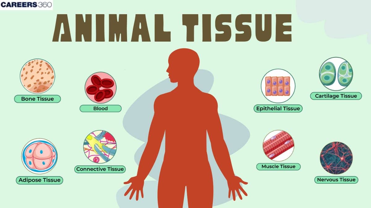
Types of Animal Tissue
There are four types of animal tissue :
Epithelial Tissue
Muscle Tissue
Connective Tissue
Neural Tissue
An animal tissue diagram typically illustrates the four main types of animal tissues: epithelial, connective, muscular, and nervous tissue, highlighting their unique structures and functions. The animal tissue diagram helps students visualize muscle tissue types, including skeletal, cardiac, and smooth muscle, each with distinct cellular arrangements and functions. A well-labelled animal tissue diagram will also include nervous tissue, showcasing neurons and glial cells that facilitate communication within the body.
Epithelial tissue
The epithelium is the layer of cells that covers the inner or outer surface. Epithelial tissue consists of compactly arranged cells with no intercellular matrix, forming continuous layers. Junctional complexes such as desmosomes and gap junctions help hold cells together. It has power or regeneration throughout life. It is avascular. Epithelial cells rest on a basement membrane that contains the acellular, matrix protein collagen. Covers the external surface of the body and internal organs. It lines the body cavity. How it covers the outer surface of the skin, the inner lining of the mouth, the digestive tract, the nose, the lungs, etc. These are protective in nature.
Types of epithelial tissues
Epithelial Tissues are always located on the inner or outer surfaces of organs, and their functions largely depend on the exact position of their locations. These are of the following types:
Type of Epithelium | Description | Location | Functions |
Thin, flat cells; may be single-layered (simple) or multi-layered (stratified). | Esophagus, blood vessels, alveoli, mouth | Protection, absorption, secretion, and diffusion. | |
Cube-shaped cells. | Renal tubules, salivary glands, sweat glands | Secretion, absorption, and protection. | |
Columnar Epithelium | Tall, column-like cells; often layered. | Urethra, anus, mammary ducts, epiglottis | Secretion and absorption. |
Ciliated Epithelium | Columnar epithelium with cilia; helps move materials. | Lining of renal tubules, airways, trachea | Movement of materials in specific directions. |
Glandular Epithelium | Modified columnar epithelium specialized for secretion. | Sweat glands, tear glands | Secretion of various substances. |
Muscular tissue
Muscle tissue makes up the muscles in our body and can contract and relax. They are made up of muscle cells. Muscle cells are elongated fibers called muscle fibres that contain the contractile proteins actin and myosin. The contractile proteins they contain assist in contraction and relaxation, resulting in locomotion and movement.
Type of muscular tissue:
Here’s a table summarizing the characteristics, locations, and functions of the three types of muscle tissue: skeletal, smooth, and cardiac.
Type of Muscle Tissue | Characteristics | Location | Functions |
Skeletal Muscle Tissue | - Striated appearance due to sarcomeres (light and dark bands) - Multinucleated cells - Voluntary control | Attached to the skeleton (e.g., biceps, quadriceps) | Facilitates body movement and posture; generates heat. |
Smooth Muscle Tissue | - Non-striated, spindle-shaped cells - Mononucleated - Involuntary control | Walls of hollow organs (e.g., digestive tract, blood vessels) | Controls involuntary movements such as peristalsis and blood flow. |
- Striated appearance - Uninucleated or binucleated cells - Involuntary control - Connected by intercalated discs | Heart only | Pumps blood throughout the body; maintains rhythmic contractions. |
Connective Tissue
Connective tissue is a specialized tissue that holds body tissues together. Connective tissue consists of a small portion of cells and a large amount of extracellular material that separates the cells. Two types of cells found in connective tissue include fibrocytes (or fibroblasts) and adipocytes, which are solid cells. In addition, the extracellular material that separates cells consists of three types of fibres: collagen fibres, reticular fibres, and elastic fibres.
Types of connective tissue:
Here’s a table describing the types of connective tissue:
Type of Connective Tissue | Description | Components | Location | Functions |
Dense connective tissue composed of chondrocytes. | Chondrocytes, semi-rigid to flexible matrix | Joints, ears, nose | Provides support and flexibility; cushions joints. | |
Hard, mineralized tissue; can be compact or spongy. | Osteoblasts, osteocytes, collagen fibers | Skeleton (e.g., femur, skull) | Supports body structure; protects organs; stores minerals. | |
Composed of fat globules; insulates the body. | Fat cells (adipocytes) | Under skin, around organs | Energy storage; insulation; cushioning. | |
Liquid connective tissue containing various cell types. | Red blood cells, white blood cells, platelets, plasma | Transports nutrients, gases, and waste products. | ||
Elastic | Contains elastic fibers; provides flexibility and support. | Chondrocytes, elastic fibers | Large blood vessels, lungs | Maintains blood pressure; aids in exhalation. |
Fibrous | Provides strength and stiffness; composed of dense fibers. | Fibroblasts, fibrous fibers | Dermis of skin, tendons | Resists mechanical stress; supports joint movement. |
Nervous tissue
Nervous tissues are made up of neurons (nerve cells in the brain), and these tissues form the entire nervous system, including the spinal cord and the brain.
Types of nervous tissue:
Here’s a table summarizing the types of nervous tissue based on the provided information:
Type of Nervous Tissue | Description | Components | Functions |
The functional unit of nerve tissue; specialized for impulse transmission. | Cytons (cell bodies), axons, dendrites | Relays nerve impulses throughout the body. | |
Non-neuronal supporting cells that assist and protect neurons. | Various types (e.g., astrocytes, microglia) | Support, nourish, and protect neurons; involved in immune response. | |
Astrocytes | Star-shaped glial cells; are most abundant in the CNS. | Radiating processes | Support neurons, control chemical environment, and guide neuron migration. |
Microglial Cells | Small, ovoid cells with thorny processes; act as immune cells in the CNS. | Phagocytic macrophages | Clean up debris and respond to injury or infection. |
Ependymal Cells | Ciliated cells lining the central cavities of the brain and spinal cord. | Ciliated epithelial cells | Form barriers between cerebrospinal fluid and CNS tissue. |
Oligodendrocytes | Cells that produce myelin sheaths around axons in the CNS. | Myelin-forming cells | Insulate axons to increase impulse transmission speed. |
Satellite Cells | Surround neuron cell bodies in the PNS; similar to astrocytes in function. | Glial cells | Support and protect neuron cell bodies in ganglia. |
Schwann Cells | Form myelin sheaths around axons in the PNS. | Myelin-forming cells | Insulate nerve fibers and support regeneration of peripheral nerves. |
Difference Between Plant Tissue and Animal Tissue
The difference between plant tissue and animal tissue lies in their structure, function, and types, reflecting the distinct roles they play in their respective organisms.
Aspect | Plant Tissue | Animal Tissue |
Cell Structure | Cells have rigid cell walls made of cellulose. | Cells lack cell walls; they have flexible membranes. |
Types of Tissue | Mainly divided into meristematic and permanent tissues. | Divided into four main types: epithelial, connective, muscle, and nervous tissue. |
Growth | Growth occurs in specific regions (meristems). | Growth is generally limited after maturity. |
Function | Primarily involved in photosynthesis, support, and storage. | Involved in movement, protection, and regulation of body functions. |
High regenerative capacity; can regenerate lost parts. | Limited regeneration capacity; healing is slower. |
Animal Tissue Culture Techniques
Animal tissue culture techniques are essential for studying cell behaviour, developing vaccines, and producing therapeutic proteins. They are vital tools in modern biological research, enabling scientists to explore cellular processes and develop new therapies. Below is a table summarizing key techniques used in animal tissue culture:
Technique | Description | Applications |
Primary Cell Culture | Involves isolating cells directly from animal tissues and culturing them. | Used for studying specific cell types and their functions. |
Cell Line Culture | Established from primary cultures; these cells can proliferate indefinitely under controlled conditions. | Useful for long-term studies and drug testing. |
Explant Culture | Small pieces of tissue are cultured to allow cells to migrate and grow in vitro. | Employed for regenerative medicine and cancer research. |
Organ Culture | Maintains whole organs in vitro to study their function and response to treatments. | Important for understanding organ-specific diseases. |
Stem Cell Culture | Culturing undifferentiated stem cells that can differentiate into various cell types. | Key for regenerative medicine and developmental biology research. |
Tips, Tricks, & Strategies to Study Animal Tissue
To effectively study the topic of Animal Tissue: Structure & Types, consider these strategies:
Tips and Tricks | Mnemonic |
Understand the four main types of animal tissues and their functions. | E.C.M.N. (Epithelial, Connective, Muscle, Nervous) |
Use diagrams to visualize tissue structures and locations in the body. | DREAM (Diagrams Reinforce Easy Analysis of Mechanisms) |
Create flashcards for key terms and examples of each tissue type. | FLASH (Flashcards Lead to Active Study Habits) |
Engage in group discussions to clarify concepts and share knowledge. | TEAM (Together Everyone Achieves More) |
Exam Weightage & Types of Questions
Knowing the weightage of this topic helps prioritize your study efforts. Here’s how it is weighted across various entrance exams:
Entrance Exam | Weightage (%) | Types of Questions |
5-10% | Conceptual questions, identification tasks | |
3-5% | Multiple-choice questions, diagram-based queries | |
4-6% | Analytical problems, comparative analysis | |
6-8% | Short answer questions, application-based queries | |
2-4% | Descriptive questions, analytical essays |
Animal Tissue NEET MCQs
Q1. The multiple layered epithelial cells are called _____.
Simple epithelial tissue
Stratified epithelial tissue
Multi-layered epithelial tissue
None of the above
Correct answer: 2) Stratified epithelial tissue
Explanation:
The multiple-layered epithelial tissue is known as stratified epithelial tissue. It consists of several layers of cells, protecting against mechanical and chemical stress. The cells in the deepest layer divide and push the older cells upward, where they may become flattened or specialized. Stratified epithelial tissue is commonly found in areas subject to wear and tear, such as the skin, mouth, oesophagus, and vagina.
Hence the correct answer is option 2) Stratified epithelial tissue.
Q2. Which of the following is a type of fluid connective tissue?
Blood
Lymph
Both a and b
None of the above
Correct answer: 3) Both a and b
Explanation:
Fluid connective tissue includes blood and lymph, both of which play essential roles in transportation and immunity. Blood consists of plasma, red blood cells (RBCs) for oxygen transport, white blood cells (WBCs) for immune defence, and platelets for clotting. It circulates nutrients, gases, hormones, and waste throughout the body. Lymph, derived from interstitial fluid, contains WBCs, particularly lymphocytes, and is crucial for immune responses, maintaining fluid balance, and transporting fats from the digestive system. Both blood and lymph serve as vital fluid connective tissues that help maintain homeostasis in the body.
Hence the correct answer is option 3) Both a and b
Q3. ________ joins muscles to bones and is made up of thick fibrous (collagen).
Areolar tissue
Tendons
Ligaments
Adipose Tissue
Correct answer: 2) Tendons
Explanation:
Tendons connect muscles to bones and are composed of thick, fibrous collagen. These structures are strong and flexible, allowing for the effective transfer of force from muscles to bones, which facilitates movement. Tendons are designed to withstand tension and provide stability to joints during physical activity.
Hence, the correct answer is option is 2) Tendons join muscles to bones and are made up of thick fibrous (collagen).
Also Read:
FAQs on Animal Tissue
What do you mean by animal tissue?
Animal tissue is a collection of specialized cells that are similar in structure and work together to perform a specific function. These tissues form the structural and functional framework of the animal body. They help in carrying out activities such as protection, movement, coordination, and transport of materials. Unlike plants, animals do not have cell walls, so tissues are more specialized for flexibility and efficiency.
What are the four types of animal tissue?
The four main types of animal tissues are:
Epithelial tissue – covers body surfaces and lines organs, providing protection and absorption.
Connective tissue – supports, binds, and connects different body parts (e.g., bone, blood, cartilage).
Muscular tissue – responsible for movement, both voluntary (skeletal) and involuntary (smooth and cardiac).
Nervous tissue – made of neurons, it controls and coordinates body functions by transmitting impulses.
What is the difference between plant and animal tissue?
Plant and animal tissues differ in structure and function. Plant tissues have a rigid cell wall and include meristematic (for growth) and permanent tissues. They focus mainly on photosynthesis, storage, and mechanical support. Animal tissues lack cell walls, allowing more flexibility. They are divided into four main types – epithelial, connective, muscular, and nervous – which help in protection, movement, coordination, and transport. Thus, animal tissues are more specialized for dynamic functions.
What are animal tissue culture techniques?
Animal tissue culture refers to the in vitro (outside the body) maintenance and growth of animal cells under controlled laboratory conditions. These techniques are widely used in medicine, genetics, cancer research, and drug testing. The major techniques include:
Primary culture: directly using tissues or cells from an animal.
Cell line culture: using already established cell lines for research.
Organ culture: maintaining the whole or part of an organ outside the body.
Stem cell culture: growing undifferentiated cells for medical research.
Frequently Asked Questions (FAQs)
Supporting tissues in animals primarily consist of connective tissues, which provide structural support, bind other tissues, and facilitate communication within the body.
The main types of connective tissue include:
- Loose Connective Tissue: Provides flexibility and support.
- Dense Connective Tissue: Includes tendons (attach muscles to bones) and ligaments (connect bones).
- Adipose Tissue: Stores fat and insulates the body.
- Cartilage: Offers flexible support in joints.
- Bone: Provides rigid structure and strength.
- Blood: Transports nutrients and waste throughout the body.
The extracellular matrix (ECM) is a network of proteins and glycoproteins that provides structural support to connective tissues and facilitates cellular functions such as growth and repair.