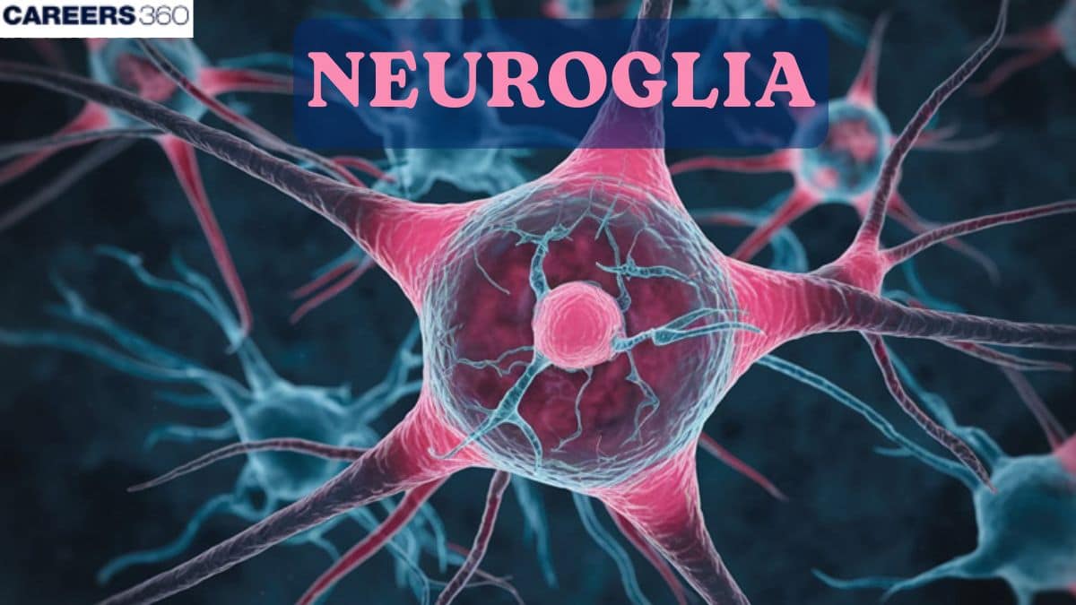
Neuroglia
Neuroglia, or glial cells, are non-neuronal cells that support, protect, and nourish neurons in the CNS and PNS. They include astrocytes, oligodendrocytes, microglia, and ependymal cells, each with vital roles in myelination, immunity, and homeostasis. A key NEET and Class 11 Biology concept.
This Story also Contains
- What are Neuroglia (Glial Cells)?
- Types of Neuroglia
- Functions of Neuroglia
- Neuroglia in Health and Disease
- Neuroglia NEET MCQs (With Answers & Explanations)
- Recommended video on "Neuroglia"
What are Neuroglia (Glial Cells)?
Neuroglia, also known as glial cells, are non-neuronal cells that surround, support, protect, and nourish neurons within both the central nervous system (CNS) and peripheral nervous system (PNS). They make up about half the volume of the CNS.
Generally, neuroglia are smaller than neurons, and they are 5 to 25 times more numerous. In contrast to neurons, glia do not generate or propagate action potentials, and they can multiply and divide in the mature nervous system. In cases of injury or disease, neuroglia multiply to fill in the spaces formerly occupied by neurons. Brain tumors derived from glia, called gliomas, tend to be highly malignant and to grow rapidly.
Types of Neuroglia
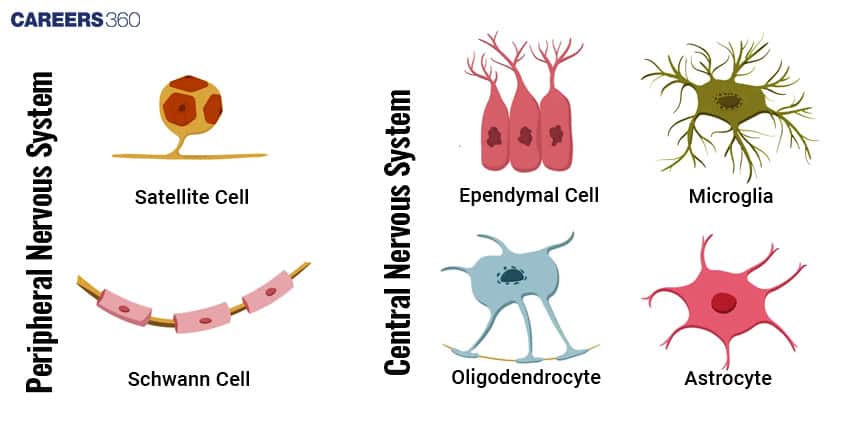
Neuroglia can be classified on the basis of size, cytoplasmic processes, and intracellular organization into four types: astrocytes, oligodendrocytes, microglial cells, and ependymal cells.
Astrocytes
These star-shaped cells have many processes and are the largest and most numerous of the neuroglia. There are two types of astrocytes. Protoplasmic astrocytes have many short branching processes and are found in gray matter. Fibrous astrocytes have many long unbranched processes and are located mainly in white matter.
The processes of astrocytes make contact with blood capillaries, neurons, and the pia mater. It provides structural support. It is also an important component of the blood-brain barrier and thus helps in maintaining the extracellular environment around neurons.
Role in Neurogenesis:
Astrocytes release growth factors and diffusible signalling molecules.
Oligodendrocytes
These resemble astrocytes but are smaller and contain fewer processes. Processes of oligodendrocytes are responsible for forming and maintaining the myelin sheath around axons. They support the rapid conduction of nerve impulses.
Impact on Nerve Impulse Conduction:
During myelination, the oligodendrocytes sheath the axon, and this causes the speed at which nerves conduct their impulses to increase because it allows electrical signals to leap from node to node of Ranvier.
Microglia
These neuroglia are small cells with slender processes that give off numerous spinelike projections. Microglial cells or microglia function as phagocytes. Like tissue macrophages, they remove cellular debris formed during normal development of the nervous system and phagocytize microbes and damaged nervous tissue.
Role in Neuroinflammation:
Microglial cells are the chief cells that participate in neuroinflammation. Thus, they respond to numerous processes of injury and disease by the exertion of positive protective and negative detrimental effects on neurological health via the release of inflammatory cytokines.
Ependymal Cells
Ependymal cells are cuboidal to columnar cells arranged in a single layer that possess microvilli and cilia. These cells line the ventricles of the brain and central canal of the spinal cord. Functionally, ependymal cells produce and assist in the circulation of cerebrospinal fluid. They also form the blood–cerebrospinal fluid barrier.
Role in CNS Homeostasis:
These cells help maintain a proper fluid balance and protective environment for the tissue of the CNS. They also assist in waste removal and provide some level of cushioning for the brain and spinal cord.
Functions of Neuroglia
The functions of neuroglia are described below-
Support and Protection
Neuroglia provide structural support to neurons and thus hold the architecture of CNS. They regulate the extra-cellular ions balance. This is very essential in the maintenance of the environment in which the neural function and synaptic activity are carried out.
Myelination and Signal Transmission
The neuroglial cells, specifically oligodendrocytes in CNS, participate in the process by which the myelin sheath is formed to enwrap the axons. This forms the basis of rapid transmission of electrical impulses along the nerve cells.
Myelination thereby vastly increases the speed and efficiency of nerve impulse transmission, which hence allows for fast communication within the CNS.
Immune Response and Neuroinflammation
Microglial cells are the chief mediators of the primary host immune response within the CNS. Thus protecting it against invading pathogens and phagocytosing dead cells and debris.
They take part in neuroinflammatory processes that, though protective in short-term responses, might be injurious in chronic conditions.
Cerebrospinal Fluid Production
Ependymal cells secrete the cerebrospinal fluid and help to circulate it, thus cushioning the brain by keeping the CNS environment stable, waste removal, and the maintenance of chemical stability.
Neuroglia in Health and Disease
The role of neuroglia are described below-
Role in Normal Functioning (Homeostasis)
Neuroglial cells are required to provide the constant environment of CNS, to assure correct activity of neurons. This control is made through the maintenance of proper ion concentrations, clearing up cell debris, and promoting synaptic plasticity.
Role in Alzheimer’s Disease
In Alzheimer's disease, changes in glial cells, represented by both astrocytes and microglia, contribute toward the stepwise events through inflammatory responses and synaptic dysfunction.
Role in Multiple Sclerosis
Destruction of oligodendrocytes, leading to loss of the myelin sheath and, consequently, disrupting the conduction of the nerve impulses, results in neurological deficits.
Neuroglia NEET MCQs (With Answers & Explanations)
Important topics that are commonly asked in NEET exam are:
Types of neuroglia & their functions
Role of microglia
Diseases linked to neuroglia.
Blood-brain barrier
Practice Questions for NEET
Q1. Which of the following cells repair the damaged neurons?
Oligodendrocytes
Astrocytes
Microglia
All of these
Correct answer: 2) Astrocytes
Explanation:
Astrocytes are star-shaped glial cells characterized by having various processes protrude from their bodies. Their function is not only to physically support the structure of nervous tissue by providing mechanical support to neurons but also to help repair damaged sites in the nervous system, hence accelerating recovery after the injury. Apart from this function, astrocytes regulate the extracellular environment and promote neuron health and functions by maintaining a balance of ions and supplying needed metabolites.
Hence, the correct answer is option 2) Astrocytes.
Q2. Which of the following cellular components is NOT present in cells that exhibit Nissl's granules and do not possess rough endoplasmic reticulum (RER) and ribosomes?
Neuroglial cells
Cells of the neural system that respond to stimuli
Dendrites of neurons
Cell bodies of excitable cells of neural tissue
Correct answer: 1) Neuroglial cells
Explanation:
Option 1 states that Neuroglial cells do not possess Nissl's granules and lack RER and Ribosomes. Neuroglial cells are supportive cells in the nervous system that provide structural and functional support to neurons. Unlike neurons, they do not exhibit Nissl's granules, clusters of RER, and ribosomes involved in protein synthesis. Neuroglial cells have different functions in the nervous systems in the nervous system, such as immune defence. Therefore, option 1 correctly identifies that neuroglial cells do not possess Nissl's granules and lack RER and ribosomes.
Hence the correct answer is option 1) Neuroglial cells.
Q3. Which of the following cells form the myelin sheath around the neurons of CNS?
Schwann cells
Oligodendrocytes
Both a and b
None of these
Correct answer: 2) Oligodendrocytes
Explanation:
Neuroglia is what sustains the neuron, and there are two major kinds, which are both macroglia: microglia is one end.
Macroglia includes
Astrocytes: They are star-shaped cells that provide mechanical support and facilitate tissue repair.
Oligodendrocytes: Tree-like cells that make myelin sheaths around CNS axons, enhancing signal transmission.
Microglia are small, phagocytic cells derived from mesoderm that function as immune cells: they engulf debris and pathogens to maintain CNS health.
Hence, the correct answer is option 2) Oligodendrocytes.
Also Read:
Recommended video on "Neuroglia"
Frequently Asked Questions (FAQs)
They act in wrapping myelin sheath around axons in the central nervous system. This wrapping allows for much quicker and more effective transmission of electrical impulses.
Microglia cells are the immune cells of the CNS. The activation of these cells, which may result from different types of injuries and diseases, can lead to the release of inflammatory cytokines which may ultimately protect the brain, but lead to its chronic inflammation and neurodegenerative disorders.
Cerebrospinal fluid works to cushion the brain, eliminate waste products, supply a stable chemical environment, and help maintain enough pressure to drive the exchange of nutrients between brain tissue and blood.
The groups of the neuroglia that provide support and protection of the neurons, maintain the extracellular environment for them, produce the myelin sheaths, contribute to immune defence, and generate the cerebrospinal fluid also known as CSF.
They include providing the blood-brain barrier that restricts the entry of large molecules, microorganisms, and viruses from the blood to the brain, structural support framework for neurons, rivers of the extracellular maintaining balanced ionic such as potassium and sodium, and secretion.