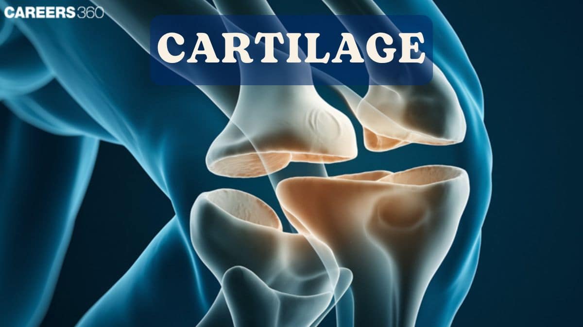
Cartilage
Cartilage is a specialized connective tissue that provides support, flexibility, and cushioning in the body. There are three main types of cartilage—hyaline, elastic, and fibrocartilage—each with distinct structure and function. Understanding its types, structure, growth, functions, and related disorders is an important concept in NEET and Class 11 Biology, as it links anatomy with movement and development.
This Story also Contains
- What is Cartilage?
- Structure of Cartilage
- Development and Growth of Cartilage
- Functions of Cartilage
- Cartilage Damage And Disorders
- Cartilage NEET MCQs (With Answers & Explanations)
- Recommended Video On 'Cartilage'

What is Cartilage?
A flexible, semifirm connective tissue inhabiting different parts of the body, cartilage serves to support, cushion, and act as a shock absorber in articulations. Cartilage is part of several anatomical structures.
There are three general types of cartilage, each with particular features and functions.
Hyaline Cartilage (Structure, Location, Functions)
It is the most common type of cartilage in the body and is recognised by the smooth, glassy appearance it bears.
Description and Characteristics
Translucent and glassy texture.
Fine collagen fibres in composition.
Provide a smooth surface for joints to move on.
Locations in the Body
Nasal septum
Trachea
Larynx
Articular surfaces of bones (joints)
Functions and Importance
Reduce friction in joints
Support and reinforcement of structure
Provides for a template of bone growth during the development
Elastic Cartilage (Structure, Location, Functions)
Elastic cartilage is adaptable and resilient due to the abundance of elastic fibres.
Description and Characteristics
Yellowish colour due to elastic fibres.
Very resilient and flexible.
Both collagen and elastic fibres are present.
Location in the Body
External ear (auricle)
Epiglottis (part of the larynx)
Functions and Importance
Provides flexible support to structures
Maintains shape but allows for movement
Protects the airway during deglutition
Fibrocartilage (Structure, Location, Functions)
Fibrocartilage is the most resilient type of cartilage and thus offers tensile strength and durability.
Description and Characteristics
Dense, fibrous texture
Contains thick bundles of collagen fibers
Resists compression and absorbs shock
Locations in the Body
Intervertebral discs (between vertebrae)
Menisci (knee joints)
Pubic symphysis (joining the pelvic bones)
Functions and Significance
Absorbs shock and reduces friction.
Supports the weight of the body against gravity in the vertebral column and lower limb bones.
Structure of Cartilage
Cartilage is a connective tissue consisting of cells embedded in an extracellular matrix that is dense and possesses elastic and viscoelastic properties.
There are several cell types in cartilage. The immature cells of the cartilage produce the extracellular matrix. Cells undergo continuous division and synthesis of the matrix components
Chondroblasts
Immature cells derived from mesenchymal cells that produce extracellular matrix.
They eventually differentiate into chondrocytes.
Chondrocytes
Mature cartilage cells are embedded in the matrix.
Maintenances of the cartilage matrix and controls of matrix composition.
Chondroclasts
Cells responsible for the resorption of cartilage
Assists in remodelling of cartilage during growth and repair
Extracellular Matrix (Collagen, Proteoglycans, Water)
The extracellular matrix forms an important component of the cartilage. It provides support to the structure by rendering it elasticity.
Structure
It contains collagen fibres which gives the tensile strength
The proteoglycans attract and hold large amounts of water, thus aiding in shock absorption.
The water contributes elasticity and spring to the matrix
Functions of ECM
Gives structure to cartilage and resists compression
Gives elasticity, thus allowing for smooth movement at joints
Allows for the exchange of nutrients and wastes through its gel nature
Cartilage development and growth occur via a series of specific biological steps.
Development and Growth of Cartilage
Cartilage develops and grows through specific biological processes.
Chondrogenesis (Embryonic Development)
Chondrogenesis is the process by which cartilage forms during development.
Mesenchymal stem cells differentiate into chondroblasts during embryonic development
Chondroblasts produce extracellular matrix and differentiate into chondrocytes
A cartilage model is formed that matures into cartilage
Cartilage increases in mass through growth and expands via specific mechanisms.
Appositional Growth
Occurs at the surface of existing cartilage
Chondroblasts in the perichondrium produce new cartilage matrix
Increases in thickness
Interstitial Growth
Takes place within the cartilage tissue.
Chondrocytes divide and produce matrices and expand cartilage from within.
Helps to increase the length of cartilage during development.
Functions of Cartilage
Cartilage performs many essential functions in the body.
Support and Shape Maintenance
Maintains the shape and structure of many body parts.
Provides structural support for flexible areas such as the ear and nose.
Protection and Joint Mobility
Cushions joints and reduces friction at joint surfaces as a result of movement.
Allows for smooth and efficient movement between joint surfaces.
Growth and Development of Bones
Part of the growth plates of long bones.
Provides support for bone growth and elongation during development.
Cartilage Damage And Disorders
Cartilage is susceptible to disorders and injuries. Various conditions are capable of causing damage to cartilage and affecting its normal function.
Osteoarthritis
Degenerative joint disease is characterised by the wearing down of cartilage.
Symptoms include joint pain, stiffness and reduced mobility.
Rheumatoid Arthritis
This type of arthritis is an autoimmune disorder causing inflammation and hence damage to the cartilage.
Causes pain in the joints, swelling and finally deformity of joints.
Chondromalacia
Softening up and thus cartilage damage typically affects the knee.
Cause pain and discomfort mainly on movement.
Cartilage NEET MCQs (With Answers & Explanations)
Important topics that are commonly asked in NEET exam are:
Structure of Cartilage (chondroblasts, chondrocytes, chondroclasts)
Growth and Development of cartilage
Cartilage Disorder
Practice Questions for NEET
Q1. Which of the following is Avascular?
Cartilage
Epithelial Tissue
Both 1 and 2
Bone
Correct answer: Both 1 and 2
Explanation:
Avascular refers to tissues devoid of blood vessels, relying on diffusion for nutrient and gas exchange. In our body, these include:
1. Cartilage: Without blood vessels, it obtains nutrients through diffusion from neighboring tissues.
2. Cornea: To maintain transparency for sight, it receives oxygen from tears and aqueous humor.
3. Epidermis: The skin's outer layer is avascular, requiring nutrients from the underlying dermis.
4. Lens: For clear vision, it lacks blood vessels and depends on the surrounding aqueous humor for nutrients.
These structures exemplify how the absence of a direct blood supply is a functional necessity in certain bodily components.
Hence, the correct answer is option 3) Both 1 and 2.
Q2. Tissues that is/are avascular and no nerve supply.
Cartilage
Bones
Epithelial Tissue
Tendons
Correct answer: 1) Cartilage
Explanation:
Cartilage is avascular connective tissue that lacks a direct nerve supply and contains no blood vessels. Its avascular nature means that cartilage depends on diffusion from adjacent tissues for its nutrients and oxygen, which could limit its thickness and repair capabilities. Epithelial tissue, being avascular, is still able to identify its stimulus. The difference between these two tissues is their specific functional adaptation in the body.
Hence, the correct answer is option 1) Cartilage.
Q3. Calcified cartilage is found in
skeleton of sharks
sternal parts of ribs
part of larynx
most of the embryonic skeleton
Correct answer: 1) skeleton of sharks
Explanation:
Cartilage is the soft skeletal tissue found more abundantly in vertebrate embryos because the bones of the adult skeleton are cartilaginous during the early stages. It is also found in the bodies of adult vertebrates. Calcified cartilage is found in the skeleton of sharks, frogs etc.
Hence, the correct answer is option 1) skeleton of sharks.
Also Read:
Recommended Video On 'Cartilage'
Frequently Asked Questions (FAQs)
Common disorders are osteoarthritis, rheumatoid arthritis, and chondromalacia.
It has a limited capacity to heal naturally and often requires medical intervention to heal significant injuries.
The structure, protection of the joints, and to provide smooth movement
Hyaline cartilage in the nose, trachea, and joints; elastic cartilage in the ear and epiglottis; fibrocartilage in intervertebral discs and menisci.