Joints: Definition, Anatomy, Movement, Types, Function

Joints: Definition, Anatomy, Movement, Types, Function

Irshad AnwarUpdated on 02 Jul 2025, 06:47 PM IST

A joint, also referred to as an articulation, is the connection between two or more bones in the body which allows movement and provides stability. In anatomy, a definition of a joint can include its role in facilitating motion and supporting the skeletal system. To define a joint, it is important to understand its structural and functional aspects. Joints are classified based on structure and function whether they are fibrous, cartilaginous, or synovial and whether they are immovable, slightly movable, or freely movable. Asking the question "What is a joint?" includes its vital role in providing mobility as well as maintaining the posture of the body. This is the topic of the Chapter Locomotion and Movement from Biology.

This Story also Contains

  1. What are Joints?
  2. Classification of Joints
  3. Structural Classification
  4. Functional Classification of Joints
  5. Advantages of Knowing Joints
Joints: Definition, Anatomy, Movement, Types, Function
Joints

What are Joints?

Joints are primary parts of the human body that enable any kind of movement and ensure stability. They connect the bones, making every living creature capable of moving. From gestures to challenging athletic moves, the types and functions of joints are highly relevant and must be learned while understanding human anatomy and physiology.

Also Read:

Classification of Joints

The classification of joints is given below in a detailed approach:

Structural Classification

Joints are structurally classified based on the material that holds the bones together.

Fibrous Joints

These joints are immovable and possess fibrous connective tissue that tightly connects bones. They are located at places in the body where more stability and support are needed, such as in the skull.

Cartilaginous Joints

Cartilaginous joints are joined by cartilage and allow limited movement. They include synchondroses, which are temporary and thus are a type of epiphyseal growth plate, and symphyses, which are permanent and connect bones in the pelvic girdle, such as the pubic symphysis.

Synovial Joints

Synovial joints have a relatively large range of motion compared to other forms of joints. Synovial joints, by their very nature, contain an enclosed cavity filled with synovial fluid. In such joints, one finds a wide degree of movement about the limbs and spine.

Commonly Asked Questions

Q: What are the main components of a synovial joint?
A:
The main components of a synovial joint include: articular cartilage covering the bone ends, a joint capsule, synovial fluid, ligaments, and sometimes menisci or articular discs. These components work together to provide smooth movement, stability, and shock absorption.
Q: What is the difference between a fibrous and a cartilaginous joint?
A:
Fibrous joints are connected by dense connective tissue and allow little to no movement (e.g., skull sutures). Cartilaginous joints are connected by cartilage and allow limited movement (e.g., intervertebral discs). Synovial joints, in contrast, allow for a wide range of motion.
Q: What is joint capsule and why is it important?
A:
The joint capsule is a fibrous sac that encloses a synovial joint. It's important because it helps maintain joint stability, contains synovial fluid within the joint cavity, and has nerve endings that provide proprioceptive information about joint position and movement.
Q: What is synovial membrane and what is its function?
A:
The synovial membrane is a thin, vascularized layer of connective tissue that lines the inner surface of the joint capsule in synovial joints. It produces synovial fluid, which lubricates the joint, and also removes debris from the joint cavity.
Q: How does the structure of the vertebral column allow for both stability and flexibility?
A:
The vertebral column consists of multiple vertebrae connected by intervertebral discs and facet joints. This structure provides stability through the interlocking processes of vertebrae, while the discs and joints allow for flexibility in various directions, balancing support and movement.

Functional Classification of Joints

Functional classification is focused on the degree and type of movements that are permitted. These include

Ball and Socket Joints

The Ball and Socket joints have the greatest range of motion, including rotation movements, which involve remodelling. Examples include the hip and shoulder joints.

Pivot Joints

Pivot joints are those that permit rotational motion around a central axis. An example is the joint between the atlas and axis vertebrae in the neck.

Hinge Joints

Hinge joints move in one plane, similar to the action of a door hinge. Examples are the elbow and knee joints.

Saddle Joints

Saddle joints are very flexible, for they allow movement in two planes. The wrist joint at the base of the thumb is an example of a saddle joint.

Condyloid Joints

The movement that is possible in condyloid joints includes flexion, extension, abduction, and adduction in two planes. Examples are the radiocarpal and metacarpophalangeal joints.

Gliding Joints

Gliding joints allow flat or slightly curved bones to slide against or alongside one another. They are articulated in the wrists and ankles.

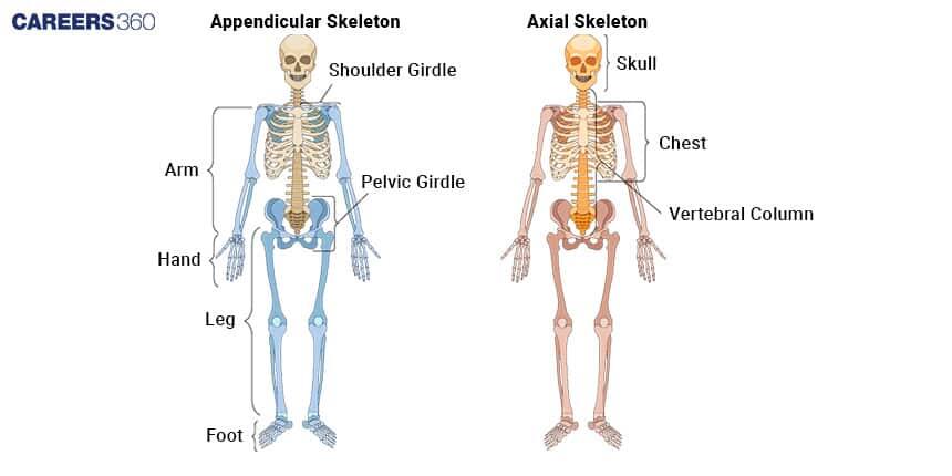
Classification of Joints
Classification of Joints

Advantages of Knowing Joints

Understanding the types and functions of joints is necessary for many aspects connected to medicine, sports science, and rehabilitation. It leads to the diagnosis and treatment of disorders of the joints, designs ergonomic tools and methods, and is effective in athletic performance.

Also Read:

Recommended video on Joints

Frequently Asked Questions (FAQs)

Q: How do the joints of the rib cage contribute to breathing mechanics?
A:
The rib cage contains several types of joints that contribute to breathing mechanics. Costovertebral joints (between ribs and vertebrae) and costotransverse joints (between ribs and transverse processes of vertebrae) allow ribs to pivot upward and outward during inhalation. The sternocostal joints (between ribs and sternum
Q: What is joint laxity and how can it impact joint function?
A:
Joint laxity refers to excessive looseness in a joint, often due to lengthened or weakened ligaments. While some degree of laxity can increase flexibility, excessive laxity can lead to joint instability, increased risk of dislocations, and potential long-term damage to joint structures. It can impact joint function by reducing stability and altering normal biomechanics.
Q: How does the temporomandibular joint (TMJ) allow for complex jaw movements?
A:
The temporomandibular joint is a modified hinge joint with unique features. It contains an articular disc that divides the joint into upper and lower compartments. This structure, along with the shape of the articulating surfaces, allows for both hinge-like movements (opening and closing the mouth) and gliding movements (side-to-side and forward-backward), enabling complex jaw motions necessary for speaking and chewing.
Q: How do condyloid joints, like those in the fingers, facilitate movement?
A:
Condyloid joints, also known as ellipsoid joints, have an oval-shaped convex surface of one bone fitting into an elliptical cavity of another. This structure, found in joints like the metacarpophalangeal joints of the fingers, allows movement in two planes - flexion/extension and abduction/adduction - enabling a wide range of finger movements.
Q: What is the role of joint mechanoreceptors?
A:
Joint mechanoreceptors are specialized sensory nerve endings found in and around joints. They detect changes in tension, pressure, and movement within the joint. This information is crucial for proprioception, motor control, and protective reflexes, helping to maintain joint stability and prevent injury.
Q: How do the sacroiliac joints provide stability while allowing subtle movement?
A:
The sacroiliac joints, between the sacrum and iliac bones, are strong, weight-bearing joints with irregular, interlocking surfaces. They provide stability for the pelvis while allowing subtle movements that are important for walking and absorbing shock during activities like running or jumping.
Q: How does the sternoclavicular joint contribute to upper limb movement?
A:
The sternoclavicular joint, where the clavicle meets the sternum, is a saddle-type synovial joint. It serves as the only bony connection between the upper limb and the axial skeleton. This joint allows the clavicle to move in multiple directions, contributing to the overall range of motion of the shoulder girdle.
Q: How do pivot joints in the neck facilitate head rotation?
A:
The primary pivot joint in the neck is the atlantoaxial joint between the first (atlas) and second (axis) cervical vertebrae. The dens of the axis acts as a pivot around which the atlas can rotate, allowing for the side-to-side rotation of the head.
Q: What is the function of the pubic symphysis joint?
A:
The pubic symphysis is a cartilaginous joint that connects the two pubic bones at the front of the pelvis. It provides stability to the pelvic girdle while allowing slight movement. In females, hormonal changes during pregnancy can increase its flexibility to accommodate childbirth.
Q: How do the joints in the foot arch contribute to shock absorption during walking?
A:
The foot contains multiple joints that form its arches. During walking, these joints, particularly in the midfoot, allow for slight movements that help distribute force and absorb shock. The flexibility of these joints, combined with the arch structure, acts like a spring to store and release energy during gait.