Synovial Joints: Definition, Meaning, Examples, Types, Diagram, Classification
Synovial joints are specialized joints in the human body that allow free movement between bones. They are characterized by a fluid-filled synovial cavity surrounded by a fibrous capsule and lined with a synovial membrane. These joints, such as the knee, shoulder, and hip, are equipped with cartilage and ligaments for stability and smooth motion. In this article, Synovial Joints, Structural Features of Synovial Joints, Types of Synovial Joints and disorders are discussed. Synovial Joint is a topic of the chapter Locomotion And Movement in Biology.
This Story also Contains
- What are Synovial Joints?
- Structural Features of Synovial Joints
- Types of Synovial Joints
- Disorders

What are Synovial Joints?
A synovial joint is one of the most paramount structural formations of the skeletal system. It plays a significant role in the elevation of movement and supports the body structure in various sections of the human structure. One of the major peculiarities is the shape of their design which allows people to move in different sections from other kinds of joints.
Structural Features of Synovial Joints
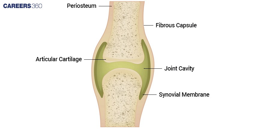
There are several key structural components of the synovial joint that are responsible for defining the functionality of the same:
Synovial Fluid
A viscous fluid fills the joint cavity lubricating the articulating surfaces and also reducing any friction inside the joint.
The Articular Capsule
It's a sheath that surrounds the joint and encloses the joint cavity, providing structural support to the joint.
Articular Cartilage
Its smooth, white tissue covers the articulating surface of the bones forming the joints. It provides frictionless, smooth motion, so the wear and tear are reduced.
Reinforcing Ligaments
Ligaments: Dense bands of fibrous connective tissue that stabilize the joint and help guide the movements of the articulating bones.
Joint Cavity
A space that exists between the articulating bones in the digits, padded by the synovial fluid enabling the free, nonbinding movement of bones.
Also Read:
Types of Synovial Joints
Six major types of synovial joints are distinguished based on the shapes of their articulating surfaces and the movements they allow:
Plane Joints
Flat articulating surfaces that allow for sliding or gliding movements.
Carpals of the wrist and tarsals of the ankle allow for movement and nonaxial playing.
Hinge Joints
Have a hinge shape configuration, thus allowing for uniaxial movement either in flexion or extension, along one plane.
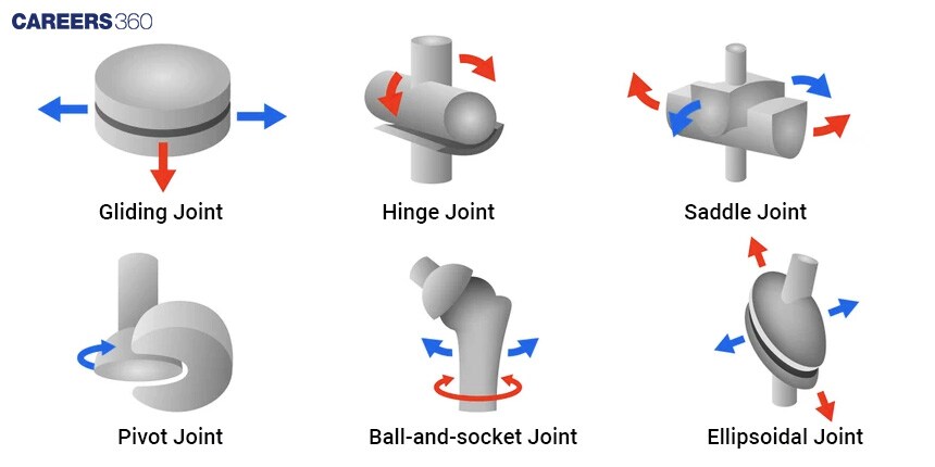
Elbow Joint
Interphalangeal joints of the fingers and toes.
Pivot Joints
The rounded end of a bone rests against another bone that has a ringlike structure, thereby allowing rotational movement along with the main axis.
A representative example is the proximal radioulnar joint, which allows the bones of the forearm, the radius, to cross over the ulna when rotating the hand from palm down to palm up positions.
Condyloid
Characterized by an oval-shaped convex surface fitting into a complementary concave surface.
Permit biaxial movements (flexion-extension and abduction adduction).
Found in the wrist joints (radiocarpal) and knuckle joints (metacarpophalangeal).
Saddle Joints
Resemble a saddle shape with concave and convex surfaces that fit together.
Allow for biaxial movements similar to condyloid joints.
Present at the carpometacarpal joint of the thumb; allows the opposition of the thumb.
Ball and Socket Joints
Ball and socket joints have the ball in one bone and a socket in another.
Ball and socket joints allow the largest range of motion of all synovial joints and also allow movement in multiple axes.
Example includes the shoulder joint and hip joint.
Disorders
Synovial joints play an extremely crucial role in movement and general body functionality. However, these joints happen to be susceptible to many disorders, such as:
Osteoarthritis: Degenerative joint disease of the articular cartilage.
Rheumatoid Arthritis: Inflammatory disease that leads to joint destruction.
Sprains and Strains: Injuries of the ligaments and tendons around a joint, leading to its stability and mobility being compromised.
Also Read:
| Ribs and Rib Cage | Vertebral Column |
| List of 206 Bones in Body | Pectoral Girdle |
| Axial Skeleton System | Appendicular Skeleton System |
Frequently Asked Questions (FAQs)
Exercising regularly, watching your pounds, and proper nutrition may keep your joints healthy and reduce the chances of having joint disorders too.
Treatment depends largely on the disorder, but it can run the gamut—from drug and exercise treatments to even joint injections or even surgery—if the case is that adverse.
Synovial joints are the joints that help perform different movements of the body, which are required to move and perform day-to-day activities.
Synovial joints encompass hinge, ball and socket, pivot, saddle, condyloid, and plane. The joints help in performing rigid movements such as bending, rotation and sliding.
Osteoarthritis, rheumatoid arthritis and joint injuries like sprains are some common disorders of synovial joints. The basic symptoms from which a person can realize the presence of such a disorder are pain, stiffness and immobility of the joint.