Centrosome: Overview, Structure, Functions, Diagram
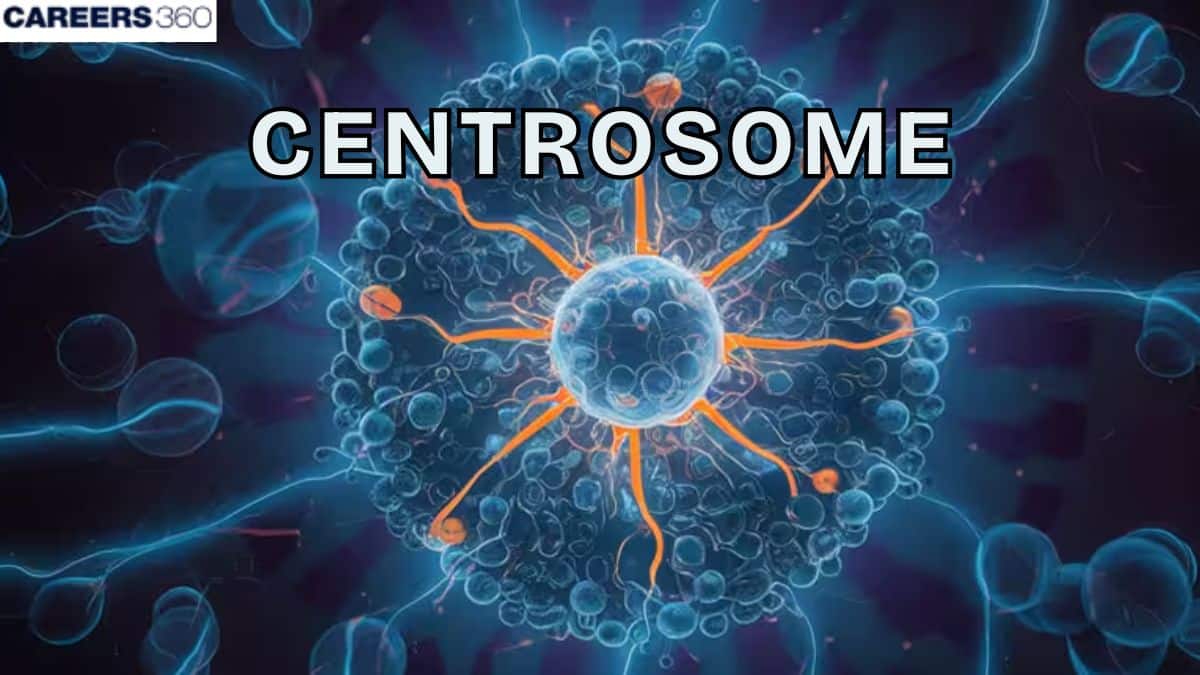
The centrosome is the primary microtubule-organizing centre (MTOC) in animal cells. It is composed of a pair of centrioles surrounded by an amorphous pericentriolar material. The centrosome plays a crucial role in cell division by forming the mitotic spindle, helps in the proper segregation of chromosomes, and regulates the cell cycle. Because of its structural and functional importance, the centrosome is a key concept in Class 12 Biology and NEET preparation.
This Story also Contains
- What is a Centrosome?
- Structure of Centrosome
- Functions of Centrosomes
- Centrosome Cycle
- Centrosomes in Different Organisms
- Centrosome Related Diseases and Disorders
- Centrosome NEET MCQs (With Answers & Explanations)
- Recommended video for Centrosome
What is a Centrosome?
The microtubule organising centre, also called the centrosome, is one of the most important structures in the cell. First identified in the 1800s by Theodor Boveri and Edouard van Beneden. It is responsible for cell shape and microtubule orientation to form a proper network and accurate cell division.
These organelles are made up of two centrioles enclosed by a matrix of proteins. They are mainly found in most of the animal cells. The role of the centrosome in organising the cell and dividing it, continues to be seen as crucial to cell biology.
Structure of Centrosome
The structure of centrosomes is given below:
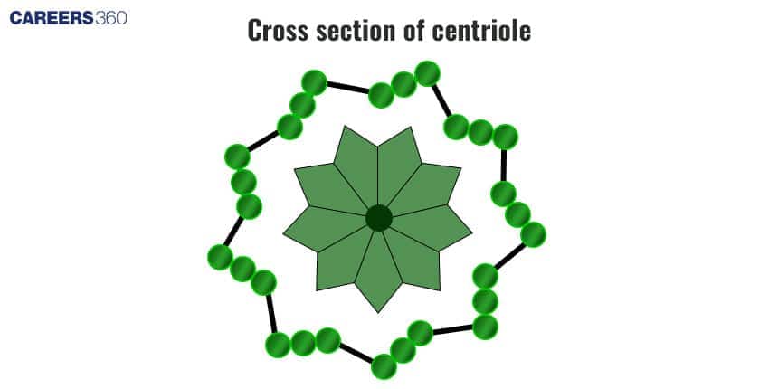
Centriole Pair (9 triplet microtubules, cartwheel structure)
A typical centrosome has two centrioles which are cylindrical structures that have nine microtubule triplets within a cartwheel-like formation. These triplets produce a frame for the formation of microtubules and are involved in the process of ciliogenesis and flagellogenesis.
Pericentriolar Material (PCM)
The centrosome matrix, also called the pericentriolar material (PCM), is a dense proteinaceous material present around the centrioles. It contains components like gamma-tubulin, pericentrin, and others, making it essential for spindle formation during cell division.
Centrosome Matrix
One of the significant components of the PCM is the centrosome matrix from which the MTOC is derived. It controls the formation and addition of microtubules and maintains the cell integrity, vehicular transport, and sometimes the mitotic spindle of the cells.
Functions of Centrosomes
The functions of centrosomes are discussed below:
Microtubule Organization and Nucleation
The centrosome is the main MTOC of animal cells organizing the network of microtubules, giving structural support and helping transport of particles. Nucleation begins at the γ-tubulin ring complexes present in the centrosome matrix, which serve as the starting sites for microtubule growth.
Role in Cell Division (mitotic spindle formation)
Centrosomes initiate the formation of new microtubules. Similarly, the centrosome also duplicates and sets as the poles of the mitotic spindle that are scattered throughout the chromatids accurately.
It also directs the formation of the mitotic spindles, which are composed of microtubes and is vital in the separation of chromosomes to the daughter cells in mitosis and meiosis.
Cell Cycle Regulation
Centrosome cycling involves its duplication and maturation, which are carefully coordinated with the cell cycle. This ensures that after cell division, each daughter cell receives one centrosome, maintaining proper cell organization and function.
Intracellular Transport
The centrosome helps in the transport of vesicles and organelles by arranging microtubules into tracks inside the cell. Along these tracks, motor proteins move vesicles and organelles to their destinations, ensuring proper functioning of the cell.
Centrosome Cycle
The centrosome cycle is discussed below:
Duplication (S-phase)
Centrosome replication takes place at the beginning of the S phase and results in the duplication of the centrosomes to two by the time the cell begins to divide. This process is controlled strictly by the proteins involved in the cell cycle like cyclins and cyclin-dependent kinases to avoid the mistakes that can cause abnormal cell division and aneuploidy.
Separation (Prophase → Metaphase)
In prophase, the centrosomes that have duplicated start to migrate and position themselves at the poles of the cell. This is led by motor proteins like the dynein and the kinesin that run along the microtubules. By the end of metaphase, the centrosomes ensure that they are located at the spindle poles where they aid in the formation of the mitotic spindle that enables the segregation of chromosomes.
Role in Cytokinesis
The centrosomes need to be positioned correctly and to function in cytokinesis. They assist in the organization of the contractile ring and the cleavage furrow that is required for the proper separation of daughter cells thereby increasing the chances of cell division success.
Centrosomes in Different Organisms
The following describes the centrosome in different organisms:
Animal Cells
The centrosomes in animal cells include two centrioles surrounded by pericentriolar material (PCM). It is the main microtubule organizing center and plays a significant role in cell division and organization. It can also manifest itself in different types of cells and at different stages of its development as a result of functional needs.
Plant Cells (no centrosomes, use other MTOCs)
Unlike animal cells, plant cells do not contain centrosomes and unipolar spindles of plant cells extend from each pole during cell division. Nonetheless, it involves other microtubule organizing centers (MTOCs). They are analogous to centrosomes and help in the organization of microtubules as well as proper cell division. The nuclear surface is usually a site of microtubule nucleation during mitosis.
Yeast and Other Eukaryotes (spindle pole bodies)
Yeast cells do not contain centrosomes. Their MTOCs are known as the spindle pole bodies (SPBs). Thus, in other eukaryotes, structurally and functionally similar organelles may be present. Such adaptations show how different species provide different solutions to the problems of assembling microtubules and dividing the cell.
Centrosome Related Diseases and Disorders
The disorders are discussed below:
Cancer
Centrosome hyperplasia, the state in which cells contain more than two centrosomes, is manifested in cancer very frequently. It can cause defects in spindle formation, chromosome segregation and antisepsis which are responsible for tumorigenesis and cancer development.
Microcephaly
The improper regulation of genes involved in centrosome duplication and function, can lead to microcephaly, a situation where an individual has a small brain. These mutations interfere with cell division and the development of the brain which in turn results in serious neurological complications.
Ciliopathies
Abnormality of the centrosome can also cause other diseases including ciliopathies. Since centrosomes are involved in forming and functioning of cilia. If proteins required for cilia do not work properly, it can result in diseases like polycystic kidney disease, respiratory diseases, and retinal degeneration.
Centrosome NEET MCQs (With Answers & Explanations)
Important topics for NEET are:
Structure of Centrosome
Functions of Centrosomes
Centrosome in different organisms
Practice Questions for NEET
Q1. When the centromere is situated in the middle of two equal arms of chromosome, the chromosome is referred as:
Metacentric
Sub-metacentric
Acrocentric
Telocentric
Correct answer: 1) Metacentric
Explanation:
When the centromere is situated in the middle of two equal arms of the chromosome, it is called a Metacentric chromosome. When the centromere is present slightly away from the middle, it is called a sub-metacentric chromosome. When the centromere is present very close to one end of the chromosome, it is known as an acrocentric chromosome. When the centromere is present at the terminal position, the chromosome is referred to as telocentric.
Hence, the correct answer is option 1) Metacentric
Q2. This cell organelle consists of two granule-like centrioles and is found in animal cells only. It helps in cell division. What is it called?
Centrosome
Chromosome
Centromere
Chromatids
Correct answer: 1) Centrosome
Explanation:
The centrosome is an organelle and microtubules are organized in the centrosome. A centrosome is composed of two centrioles, mother centrioles, and daughter centrioles which are arranged perpendicular to each other. They are structures found inside of cells, centrioles are microtubule rings. They pull chromatids about during cell division.
Hence, the correct answer is option 1) Centrosome.
Q3. The major function of centrioles is
Cell division
Cell metabolism
Cell respiration
All of these
Correct answer: 1) Cell division
Explanation:
The major function of centrioles is to organize the microtubules during cell division, specifically in the formation of the spindle fibres. These spindle fibres help separate the chromosomes to opposite poles of the cell during mitosis and meiosis. Centrioles are located in the centrosome, which acts as the microtubule organizing centre (MTOC). They play a key role in maintaining the cell's shape and structure by anchoring microtubules. In addition, centrioles are involved in the formation of cilia and flagella, which are essential for cell movement and fluid movement across surfaces. Centrioles duplicate during the cell cycle to ensure that each daughter cell has the necessary components for proper division.
Hence, the correct answer is option 1) Cell division
Also Read:
Recommended video for Centrosome
Frequently Asked Questions (FAQs)
Some of the diseases related to centrosomes include cancer, microcephaly and ciliopathies.
The main structures that form a centrosome include two centrioles; they are cylindrical structures that consist of microtubule triplets. Around the centrioles, there is the pericentriolar material or PCM, which is a proteinaceous matrix composed of various proteins.
Centrosome replication happens during the S phase. Centrosome duplicates and the two centrosomes are pulled apart and move to the proximal poles of the cell during mitosis. This process is strictly controlled to achieve the correct course of the formation and operation of the mitotic spindle.
The centrosome in a cell is mostly involved with the manipulation of microtubules and the management of a cell cycle. It is the principal structure required for nucleation of microtubules necessary in the maintenance of cell form, movement of organelles and objects within the cell, and information of the spindle during mitosis.