Cilia
Cilia are tiny hair-like projections on the surface of eukaryotic cells, made of microtubules in a “9+2” or “9+0” arrangement. They help in locomotion, movement of fluids, and sensory functions. Cilia are essential in processes like respiration, reproduction, and signal transduction, making them an important topic in Cell: The Unit of Life for NEET and Class 11 Biology.
This Story also Contains
- What is Cilia?
- Types of Cilia
- Structure of Cilia
- Functions of Cilia
- Cilia vs Flagella (Comparison Table)
- Cilia in Different Organisms and Systems
- Cilia Disorders (Ciliopathies)
- Cilia NEET MCQs (With Answers & Explanations)
- Recommended video for Cilia
What is Cilia?
Cilia are short, bristle-like structures that arise from the surface of eukaryotic cells and have numerous functions, including motility and sensation. These organelles are involved in many biological activities, such as the migration of cells, the movement of fluids and particles over the cell’s surface and signal transduction.
The arrangement of cilia is comparable to eukaryotic flagella, an arrangement 9+2 containing microtubules where the axoneme is surrounded by the cell membrane. Cilia can be a locomotor, allowing movement of fluids in respiratory tracts or oviducts, or non-locomotor, also known as primary cilia, which are involved in sensory functions.
Types of Cilia
There are two types of cilia based on their locomotory ability:
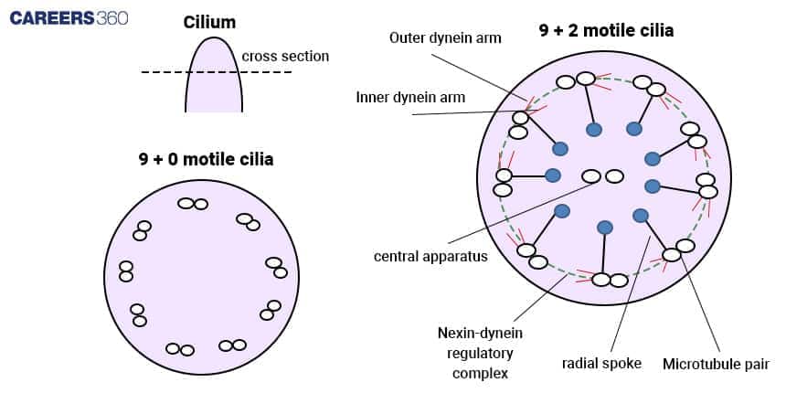
Motile Cilia (9+2 Arrangement)
The motile cilia are made of the axoneme containing a specific pattern called “9+2” microtubules where nine doublets are embraced centrally by a pair. They are connected with the cell by the basal body which is somewhat similar to the centriole in structure.
Function in Movement: These cilia move in a coordinated manner, or rather beat in a wave-like motion to result in movement. This action is necessary for functions such as mucus and debris movement in the respiratory tracts and pushing eggs along the oviducts.
Non-motile (Primary) Cilia (9+0 Arrangement)
Primary cilia possess a “9+0” microtubular arrangement in the axoneme and do not have the central pair that is seen in motile cilia. They are also inserted on a basal body but they are often shorter in size and number as compared to motile cilia.
Function as Sensory Organelle: While motile cilia have a function in movement, primary cilia do not. Primary cilia act as mechanosensitive and chemosensitive organelles. They collect information from the cell's environment and are involved in signaling pathways that regulate cell division and specialization.
Structure of Cilia
The cilia structure and composition are given below:
Axoneme
"9+2" microtubule arrangement in motile cilia:
Motile cilia have a different axoneme structure known as “9+2”. This is a ring composed of nine sets of microtubules and two central microtubules. It is arranged this way for the rhythmic and synchronised beating of motile cilia in a waving kind of method.
"9+0" microtubule arrangement in primary cilia:
Primary or nonmotile cilia have a ‘9+0’ structure implying that while there are nine microtubules that form nine detachments, there are no microtubules in the centre. It is in this respect that the structure supports their function as sensory organelles and not for movement.
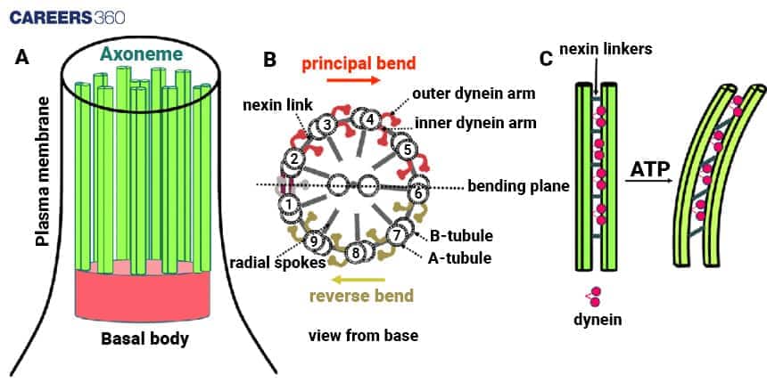
Basal Body
A cilium is built from microtubules that are nucleated at the basal body. The structure resembles a centriole but its function is to anchor the cilium to the cell membrane.
Astral is involved in arranging microtubules into the axoneme and supports the emergence of the cilium from the cell membrane.
Microtubules and Tubulin Proteins
Microtubules consist of tubulin proteins, which includes alpha-tubulin and beta-tubulin, and are present in motile and primary cilia. These proteins form a coiled structure that creates the cylindrical shape of cilia. This provides support and stability to the organelle.
Dynein Arms and ATP
Dynein arms are membrane-bound proteins anchored to ciliary microtubule doublets in organisms that bear cilia. They utilize ATP as an energy source to slide the microtubules against each other. This sliding movement causes the cilium to bend and when repeated in coordinated manner, they produce wave-like beating motion that enables movement.
Functions of Cilia
The cilia function and mechanism is listed below-
Locomotion in Protists (e.g., Paramecium)
In single-celled organisms such as Paramecium, the motile cilia form all over the cell surface and undulate in a coordinated fashion to enable swimming in water. Thus, this kind of movement enables Paramecium to move throughout its milieu, escape the jaws of its predators and find its food.
Fluid Movement in Respiratory Tract
In complex organisms, the motile cilia are vital in moving fluid over an epithelial layer. For instance, in the human respiratory tract, cilia have a rhythmic beating motion that is involved in the movement of mucus containing dust and pathogens out of the lungs and towards the throat so that they can be either coughed out or swallowed.
Sensory Functions and Signal Transduction
Primary cilia are involved in mechanosensation, that is, they control the reception of signals from the environment and their transduction to the cell. They are involved in several signalling pathways including the Gli protein signalling, or the Hedgehog protein signalling, involved in cell differentiation and organ development. Any malfunction in the primary cilia can alter their role in cellular signalling and structure.
Cilia vs Flagella (Comparison Table)
Although cilia and flagella are used in locomotion, they differ in their structure, function and composition. This tables gives the major differences between the two:
Feature | Cilia | Flagella |
Length | Shorter (5-10 µm) | Longer (10-200 µm) |
Number per Cell | Numerous, often hundreds per cell | Few, typically 1-8 per cell |
Movement | Coordinated, wave-like beating | Rotational (prokaryotes) or wave-like (eukaryotes) |
Microtubule Arrangement | "9+2" in motile cilia, "9+0" in primary cilia | "9+2" in eukaryotic flagella, different in prokaryotes |
Function | Locomotion, fluid movement, sensory roles | Locomotion, sometimes sensory functions |
Examples | Respiratory tract cilia, cilia in Paramecium | Sperm flagella, bacterial flagella |
Anchoring Structure | Basal body | Basal body (eukaryotes), motor complex (prokaryotes) |
Energy Source | ATP (via dynein arms) | ATP (eukaryotes), proton motive force (prokaryotes) |
Presence in Cells | Found in eukaryotic cells | Found in both prokaryotic and eukaryotic cells |
Beating Pattern | Synchronised, often metachronal rhythm | Unsynchronised, propeller-like (prokaryotes), or wave-like (eukaryotes) |
Cilia in Different Organisms and Systems
Cilia in different organisms and systems are listed below:
Protists
In the genera of protists including Paramecium and Tetrahymena, the cilia are distributed all over the cell body and are involved in movement and feeding. These cilia move in cyclic motions and enable the organisms to swim in water and guide food particles towards the oral groove for consumption.
Sensory Functions in Kidney & Eye
The primary cilia are immotile and have an important part in sensory functions in several organs and tissues. In kidneys, primary cilia sense the flow of fluid and respond to maintain the functioning of the kidney. In eyes, they are involved in photoreceptor cells which are vital in the process of sensing light and vision.
Human Reproductive System
In the female reproductive system, cilia embedded in the lining of the fallopian tubes assist in moving the egg from the ovary to the uterus. In the male reproductive system, cilia help to move sperms in their way in the reproductive system to have fertilization.
Human Respiratory System
In the human respiratory tract, the epithelial cells are covered by motile cilia which act as a transport machinery to move mucus and trapped particles from the lungs. The coordinated beating of cilia carries the mucus in an upward fashion towards the throat, effectively trapping and removing pathogens in the airways.
Cilia Disorders (Ciliopathies)
Ciliopathies are associated with malfunctioning cilia and there exist various diseases associated with the improper functioning of cilia.
Primary Ciliary Dyskinesia
Primary ciliary dyskinesia is a disorder where movements of cilia are reduced and leads to recurrent chest infections, reduced fertility and hearing difficulty.
Polycystic Kidney Disease (PKD)
Polycystic Kidney Disease (PKD) is associated with defects in primary cilia within the cells of the kidney. This results in the formation of cysts that are filled with fluid, and the end outcome is renal failure.
Chronic Bronchitis
In chronic bronchitis, a person experiences loss of cilia which leads to thick build-up of mucus and repeated infections. If the cilia that line the bronchial tubes get damaged, they cannot help to move the mucus out. Thus leading to obstruction of the airway and inflammation.
Cystic Fibrosis
In cystic fibrosis, mucus is tenacious, thick and effective to ciliary movement that results in severe disorders affecting the respiratory and digestive system due to mucus deposition and high incidence of infections.
Cilia NEET MCQs (With Answers & Explanations)
Important topics for NEET are:
Types of Cilia (Motile and Non-motile)
Cilia vs Flagelle
Cilia in different organisms
Disorders related to Cilia
Practice Questions for NEET
Q1. ______ are present in large numbers on the surface of the cell.
Flagella
Cilia
Frimbriae
All of these
Correct answer: 2) Cilia
Explanation:
Cilia are short, hair-like structures that are used to move the entire cell. Many cilia extend along the entire surface of the cell.
Structure of Cilia:
Cilia and flagella have a similar structure made up of microtubules arranged in a 9 + 2 format.
At the core lie two pairs of microtubules that are encased within 9 pairs of microtubules.
Cilia are relatively shorter cellular appendages that are present over the entire surface of the cells.
Hence, the correct answer is option 2) Cilia.
Q2. The arrangement of outer and central microtubule in a cilium is called the
9+1 pattern
9+0 pattern
Flagellium pattern
9+2 pattern
Correct answer: 4) 9+2 pattern
Explanation:
The 9+2 pattern is the configuration of the core and outer microtubules in a cilium.
Nine pairs of microtubules are grouped in a circle around two solitary microtubules in the middle of this formation. The majority of eukaryotic cilia and flagella have this pattern, which is crucial to their mobility and functionality.
Hence, the correct answer is option 4) 9+2 pattern.
Q3. Select the incorrect statement regarding cilia.
Cilia is of two types
Stereocillium is found in epididymis and Vas deferens.
Organization of (9+2) found in Kinetocilium
Stereocilium 9+2 is present and is non motile.
Correct answer: 4) Stereocilium 9+2 is present and is non motile.
Explanation:
Some cells, like those in the inner ear and epididymis, have non-motile hair-like projections called stereocilia on their surface. They lack the 9+2 microtubule pattern that distinguishes motile cilia, nevertheless.
The 9+2 microtubule arrangement of motile cilia, which are present in organs like the oviduct and respiratory tract, allows them to move and carry out tasks like transferring eggs or mucus.
Stereocilia differ from cilia in that they frequently lack the 9+2 configuration, are non-motile and have a simpler internal structure.
Hence, the correct answer is option 4) Stereocilium 9+2 is present and is nonmotile.
Also Read:
Recommended video for Cilia
Frequently Asked Questions (FAQs)
Cilia are straw-like structures that are present at the outer side of the cell. They perform many tasks like the movement of an organism from one place to another, the movement of fluids, and some type of sensation.
Motile cilia is capable of moving, bending to produce a flowing motion or moving a cell through its milieu. They usually have the “9+2” organisation. Primary cilia are not capable of movement. They have the ‘9+0’ microtubule and is included in signal transduction and cellular communication.
Cilia are built from tubulin proteins that are a part of the microtubules of the axoneme, which is the main structural component of cilia. Other proteins include dynein arms, kinesin, and various motor proteins.
Cilia are involved in cell transport and moving fluids by coordination of beating or waving of cilia. The motility of cilia is coordinated where the coordinated whip-like movement produces propulsion and hence the cell can swim or move fluid over the cell surface.
Ciliopathies are a category of heterogeneous diseases originating from mutations in cilia proteins. Some examples include; primary ciliary dyskinesia, polycystic kidney disease, Bardet-Biedl syndrome, Meckel-Gruber syndrome, and Joubert syndrome.