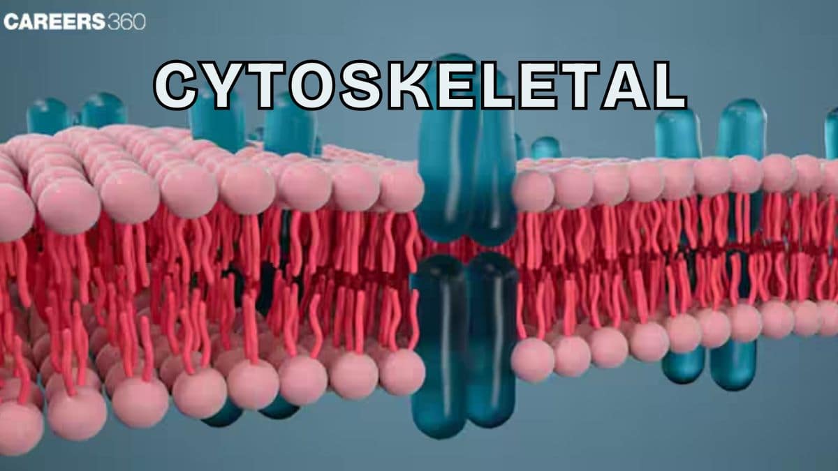
Cytoskeleton: Definition, Types, Examples, Diagram, Functions, Structure
The cytoskeleton is a dynamic network of protein filaments that maintains cell shape, enables intracellular transport, and drives processes like cell division and movement. It consists of microfilaments, intermediate filaments, and microtubules. A core NEET and Class 11 Biology concept.
This Story also Contains
- What is Cytoskeleton?
- Structure of Cytoskeleton
- Functions of Cytoskeleton
- Cytoskeleton and Motor Proteins
- Cytoskeleton and Cell Signaling
- Cytoskeleton in Disease
- Experimental Techniques to Study Cytoskeleton
- Cytoskeleton NEET MCQs (With Answers & Explanations)
- Recommended Video For Cytoskeleton
What is Cytoskeleton?
Cytoskeleton is a network of protein filaments and tubules present in the cytoplasm of eukaryotic cells. It was initially thought of as having only a role in maintaining cell shape, but its functions extend to enabling intracellular transport, facilitating cell division, and supporting cellular movements. Therefore, the cytoskeleton forms a key role in cellular biology.
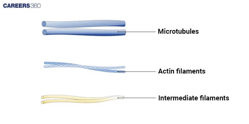
Structure of Cytoskeleton
Three types of filamentous proteins make up the cytoskeleton – actin filaments, microtubules, and intermediate filaments. Together they maintain the shape and mechanics of the cell.
Microfilaments (Actin Filaments)
These are less rigid than microtubules and the smallest in diameter (8 nm).
They are the polymers of small globular proteins called actin.
Actin is arranged in a helix that completes a turn every 13 subunits. The composition of actin depicts a bundle of fibres influenced by the dissociation rate and the type of cross-linker. These structures are formed in response to compression and tension.
F-actin protofilaments are made up of G-actin subunits. F-actin is the filamentous actin, while G-actin is the globular actin.
Actin helps in chemotaxis, cell-cell interactions, cytokinesis, and changes in cell shape during phagocytosis and muscle contraction.
They provide shape and rigidity to the cells. They can depolymerise (disassemble) and reform quickly, thus enabling a cell to change its shape and move.
Intermediate Filaments
Intermediate filaments are the least rigid, less dynamic and resist tensile forces more effectively than the compressive forces.
They are called intermediate filaments because their diameter (8 to 10 nm) is between microfilaments and microtubules.
These are structural in function and do not perform any role in the movement.
Some intermediate filaments are associated with specific cell types. For example, neurofilaments in neurons, fibroblasts in blood cells, etc.
Nuclear lamins which consist of intermediate filaments help in maintaining the integrity of the eukaryotic nucleus.
These are formed through the process of polymerization.
Microtubules
Microtubules are the largest types of filaments with a diameter of 25 nm.
Their walls are made up of polymerised dimers of a-tubulin and B-tubulin. Tubulins are constantly added and subtracted at both ends.
Microtubules are the most rigid and have complex assembly and disassembly dynamics.
During mitosis, the microtubule cytoskeleton rearranges itself into a mitotic spindle and facilitates chromosome alignment and segregation. This property of the microtubule depends on the complex assembly dynamics of individual microtubules.
Like microfilaments, microtubules can dissolve and reform quickly.
Functions of Cytoskeleton
The functions of cytoskeleton is given below-
Cell Shape and Mechanical Support
Provides a structural framework, maintaining the cell’s shape.
Actin filaments form a cortical network beneath the plasma membrane.
Intermediate filaments distribute mechanical stress across the cell.
Microtubules resist compression and help maintain cell rigidity.
Intracellular Transport
Mechanisms of transport along microtubules and actin filaments.
Vesicles and organelles are transported along microtubules via motor proteins.
Actin filaments facilitate short-range transport within the cell.
Cell Division and Movement
Cytoskeleton helps in the movement of chromosomes during cell division through spindle fibers (microtubules).
It supports the separation of daughter chromosomes to opposite poles.
Microfilaments and microtubules assist in cytoplasmic streaming and organelle movement.
Cytoskeleton and Motor Proteins
The architecture of the networks formed by cytoskeletal filaments is controlled by several types of regulatory proteins. The three main motor proteins are:
Kinesin moves cargo towards the plus end of microtubules (away from the nucleus).
Dynein moves cargo towards the minus end of microtubules (towards the nucleus).
Myosin transports cargo along actin filaments, involved in muscle contraction and various cellular processes.
Cytoskeleton and Cell Signaling
The cytoskeleton and cell signalling is discussed below-
Interaction with Cell Membrane
The cytoskeleton is linked to the plasma membrane by a variety of proteins. It is important for the maintenance of the cell shape and provides a scaffold for interactions with the outside world.
This linkage also serves as a basis for signal transduction pathways. It involves receptors in the membrane that then activate the cytoskeleton to serve in information processing by turning those signals into action, which in turn affects cellular responses.
Response to External Stimuli
The ability of cytoskeleton structures to reorganize themselves in response to mechanical or chemical signals allows the cell to adapt according to these changes. An example is the dynamic response needed during processes such as migration, division, and differentiation. It enables the cell to respond appropriately to any external stimulus it faces.
Cytoskeleton in Disease
Many diseases are associated with cytoskeletal defects. The major diseases include:
Neurodegenerative Disorders (Alzheimer’s, Parkinson’s)
Neurodegenerative diseases such as Alzheimer's and Parkinson's diseases, are diseases characterized by neuronal loss resulting from defects in microtubules and actin filaments.
Cancer and Metastasis
In cancer, mutations cause uncontrolled cell division and metastasis. Knowledge of these defects has contributed to our understanding of these disorders and may be important in developing targeted treatments.
Experimental Techniques to Study Cytoskeleton
Various experimental techniques to study the cytoskeleton are explained below:
Microscopy Techniques
Various microscopy techniques, such as fluorescence microscopy, and electron microscopy, among others, are used. These give a detailed visualisation of cytoskeletal structures.
Biochemical Approaches (Western Blot, Fractionation)
The techniques also include the fractionation and purification of proteins to isolate the cytoskeletal proteins. Western blotting is a highly sensitive biochemical method to detect proteins.
Genetic Approaches (Knockouts, RNAi)
Genetic approaches, such as gene knockouts and RNA interference (RNAi), also provide a way to identify roles for specific cytoskeletal proteins.
Cytoskeleton NEET MCQs (With Answers & Explanations)
Important topics for NEET are:
Microfilaments, Intermediate filaments, Microtubules
Functions of Cytoskeleton
Motor Proteins (Kinesin, Dynein, Myosin)
Practice Questions for NEET
Q1. Which of the following functions is carried out by the cytoskeleton in a cell?
Nuclear division
Protein Synthesis
Motility
Transportation
Correct answer: 3) Motility
Explanation:
The cytoskeleton is a network of protein filaments found in the cytoplasm of cells. It plays a crucial role in providing structural support, maintaining cell shape, and facilitating various cellular processes. One of the key functions of the cytoskeleton is cell motility, which includes the movement of cells themselves and the movement of cellular components within the cell.
The cytoskeleton is responsible for the movement of cells through processes such as amoeboid movement, ciliary/flagellar movement, and muscle contraction. It provides the necessary structure and organization for these movements to occur.
While nuclear division (Option 1) is primarily regulated by the spindle apparatus during mitosis and meiosis, and protein synthesis (Option 2) is carried out by ribosomes, the cytoskeleton is not directly involved in these specific processes.
Transportation (Option 4) can involve the cytoskeleton indirectly, as it provides tracks or highways for intracellular transport, but it is not the primary function of the cytoskeleton. The cytoskeleton's primary role in transportation is to help maintain the cellular architecture and provide support for the transport systems.
Hence, the correct answer is option 3) Motility.
Q2. Microtubules are the constituents of:
Cilia, Flagella and Peroxisomes
Spindle fibres, Centrioles and Cilia
Centrioles, Spindle fibres and Chromatin
Centrosomes, Nucleosome and Centrioles
Correct answer: 2) Spindle fibres, Centrioles and Cilia
Explanation:
Microtubules are unbranched hollow submicroscopic tubules of protein tubulin which develop on specific nucleating regions and can undergo quick growth or dissolution at their ends by assembly or disassembly of monomers. Except for Slime Moulds and Amoebae, microtubules occur widely in eukaryotic cells. They are present in the cytoplasm as well as in specialized structures like centrioles, basal bodies, cilia or flagella, sensory hair, equatorial rings of thrombocytes, spindle fibres, chromosome fibres, etc.
Hence, the correct answer is option 2) Spindle fibres, Centrioles and Cilia.
Q3. Mitotic spindle, produced during nuclear division, is mainly composed of which protein?
Actin
Tubulin
Actomyosin
Myoglobin
Correct answer: 2) Tubulin
Explanation:
The actomyosin ring is a prominent structure during cytoplasmic division. Spindle fibers are made up of a protein called tubulin. The actomyosin ring, composed of actin and myosin filaments, contracts to facilitate cytokinesis by dividing the cytoplasm during cell division. Spindle fibers, made of tubulin, play a crucial role in separating chromosomes during mitosis and meiosis. These structures ensure accurate chromosome segregation and the proper division of cellular contents into daughter cells.
Hence, the correct answer is option 4) Tubulin.
Also Read:
Recommended Video For Cytoskeleton
Frequently Asked Questions (FAQs)
Microtubules help in the movement of chromosome in the cell.
These diseases may be a result of cytoskeletal defects in the cell: neurodegenerative disorders, Alzheimer's and Parkinson's.
It is called the cytoskeleton because it provides for structural support and shape, movement and intracellular transport cell's skeleton.
Microfilaments are thin actin filaments that are involved in cell movement, intermediate filaments are formed to give mechanical strength, and microtubules are thick tubulin structures to assist in cell division and transport.
The cytoskeleton, during the process of cell division, is responsible for assembling the mitotic spindle, segregation of the chromosomes, and driving cytokinesis.