Animals Nervous System: Introduction, Classification, Spinal Cord, Neurons
The animal nervous system is a complex network of the brain, spinal cord, and nerves that coordinates and controls body activities. It processes environmental stimuli and directs responses, helping animals adapt and maintain homeostasis. Understanding its structure, types of neurons, and brain divisions is crucial for NEET and Class 11 Biology.
This Story also Contains
- What is the Nervous System?
- Structure of the Nervous System
- Neurons: Functional Units
- Neuroglia: Supporting Cells
- Cerebrospinal Nervous System
- Types of Nerves
- Parts of the Human Brain
- Animal Nervous System NEET MCQs
- FAQs on Animal Nervous System
The nervous system is a rapid-communication system that helps with the neural control and coordination of the body. This system processes information from the environment and directs appropriate responses. The basic unit of nervous integration in all animals is the neuron, a highly specialized cell designed to conduct selfpropagating electrical signals, called action potentials. Action potentials are transmitted from one neuron to another across synapses, which may be either electrical or chemical. The nervous system consists of the brain, spinal cord, and nerves, forming the central and peripheral nervous systems.
What is the Nervous System?
The nervous system is the network of nerves and cells that integrates and transmits action and signals between parts of an animal's body. It is an important part of the organ that serves a principal function in controlling and coordinating body activities, from simple to very complex, sensory perception, and maintaining homeostasis. As such, it enables animals to monitor, adapt, and react to both sets of internal and external stimulations.
Structure of the Nervous System
The broad division of the nervous system is classified into two parts: the Central Nervous System and the Peripheral Nervous System.
Central Nervous System (CNS)
The Central Nervous System consists of two major divisions:
Brain
The structure and functions of the cerebrum include higher brain functions such as thought, action, and sensory processing.
The structure and functions of the cerebellum include control over balance, coordination, and fine muscle control.
The structure and functions of the brainstem include regulating vital functions of the body such as heartbeat, breathing, and sleep cycles.
Spinal Cord
A cylindrical bundle of nerve fibres extending from the brainstem down the vertebral column.
It transmits information between the brain and the rest of the body and mediates reflex actions.
Peripheral Nervous System (PNS)
The Peripheral Nervous System has the following components:
Somatic Nervous System
It is responsible for controlling voluntary movements and transmitting sensory information towards the CNS.
This comprises nerves connecting to muscles and sensory receptors.
Autonomic Nervous System
Sympathetic Division: This prepares the body for 'fight or flight' responses.
Parasympathetic Division: Serves 'rest and digest' activities.
Neurons: Functional Units
Neurons are the simplest units of nervous systems that carry nerve impulses. Neurons (nerve cells) possess electrical excitability, the ability to respond to a stimulus and convert it into an action potential.
Functionally, neurons are classified according to the direction in which the nerve impulse is conveyed.
Sensory or afferent neurons: These either contain sensory receptors at dendrites. Once an appropriate stimulus activates a sensory receptor, the sensory neuron forms an action potential in its axon and the action potential is conveyed into the CNS through cranial or spinal nerves. Most sensory neurons are unipolar in structure.
Motor or efferent neurons: These convey action potentials away from the CNS to effectors (muscles and glands) in the periphery (PNS) through cranial or spinal nerves. Motor neurons are multipolar in structure.
Interneurons: They are mainly located within the CNS between sensory and motor neurons. Interneurons process incoming information from sensory neurons and then elicit a motor response by activating the appropriate motor neurons. Most interneurons are multipolar in structures.
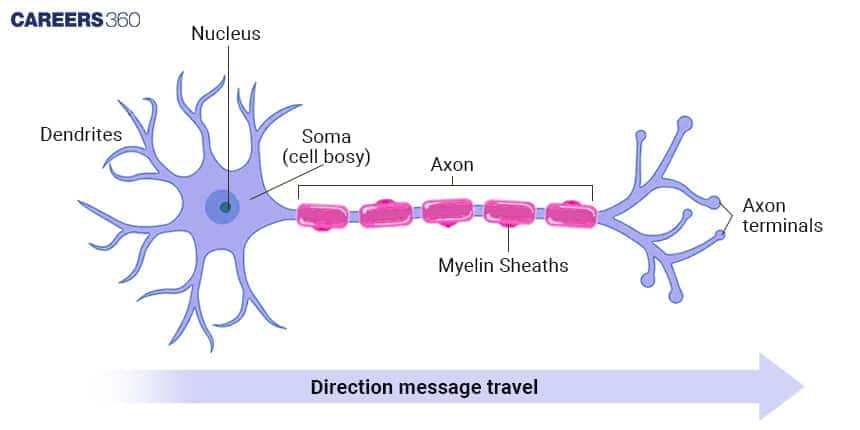
Structure:
Most neurons have three parts: a cell body, dendrites, and an axon.
Cell Body (Soma): It contains a nucleus surrounded by cytoplasm that includes typical cellular organelles such as lysosomes, mitochondria, and a Golgi complex.
Dendrite: They are the receiving or input portions of a neuron. The plasma membranes of dendrites contain numerous receptor sites for binding chemical messengers from other cells. Dendrites usually are short, tapering, and highly branched.
Axon: The single axon of a neuron propagates nerve impulses toward another neuron, a muscle fiber, or a gland cell. It is a long, thin, cylindrical projection.
Neuroglia: Supporting Cells
Astrocytes: Provide mechanical support and protection for neurons, and maintain the blood-brain barrier.
Oligodendrocytes: Generate myelin in the CNS.
Microglia: Serve as immune cells within the CNS.
Ependymal Cells: Line ventricles and produce CSF.
Cerebrospinal Nervous System
It is made up of twelve pairs of cranial nerves, each of which has a specific function and is related to the brain. The nerves and their corresponding roles are as follows:
Optic: Sight
Oculomotor: Movement of the eyeball, pupils, and lens
Olfactory: The sense of smell
Trochlear: Eye muscle movement (superior oblique)
Trigeminal: Provides nerves to the cheeks, mouth, eyes, and controls chewing
Abducens: Human lateral rectus muscle action and outward vision
Facial: controls salivary gland function, facial muscle movement, and anterior tongue taste perception
Glossopharyngeal: A gustatory experience
Acoustic: Preserves hearing and balance
Vagus: Provides the organs in the chest and belly with nerve supply
Spinal accessory: Movement of the head and shoulders
Hypoglossal: Controls the tongue's muscles
Types of Nerves
A nerve is created when many axon fibres are bundled together. There are three kinds of nerves:
Sensory: The nerve fibres are referred to as sensory when the impulse travels from the receptor to the brain or spinal cord. The nerves in the ears, eyes, and nerves, for instance.
Motor: A motor neuron is what happens when an impulse travels from the brain or spinal cord to a gland or muscle.
Mixed: Both the motor and sensory nerves are found in a mixed nerve. For instance, the spinal nerves
Parts of the Human Brain
The sense organs send messages to the human brain, which then relays those signals back to the nerves. To shield the brain from mechanical shocks, it is housed inside the skull, which also houses the cerebrospinal fluid. It is separated into three areas:
Forebrain
The receptors send the impulses to it. It contains distinct sections for analysing the various signals, such as smell, hearing, etc. This is also where the thought process occurs. After being analysed, the incoming signals are sent to the appropriate areas.
Midbrain
Numerous voluntary and involuntary processes are carried out by our bodies. Humans are able to manage voluntary movements like pushing and running. Blinking and breathing are examples of involuntary behaviours that are automatic and beyond human control.
Hindbrain
The cerebellum and medulla are located in the hindbrain. These regulate blood, saliva, and respiration.
Animal Nervous System NEET MCQs
Q1. The somatic neural system
is the site of information processing and control.
relays impulses from the CNS to skeletal muscles
is further classified into the sympathetic neural system and parasympathetic neural system.
transmits impulses from the CNS to the involuntary organs and smooth muscles of the body.
Correct answer: 2) relays impulses from the CNS to skeletal muscles
Explanation:
Human Neural System:
The PNS is divided into two divisions called the somatic neural system and the autonomic neural system.
The somatic neural system relays impulses from the CNS to skeletal muscles.
The autonomic neural system transmits impulses from the CNS to the involuntary organs and smooth muscles of the body.
The autonomic neural system is further classified into the sympathetic neural system and parasympathetic neural system.
The somatic neural system relays impulses from the CNS to skeletal muscles.
Hence, the correct answer is option 2) relays impulses from the CNS to skeletal muscles.
Q2. Myelinated nerve fibres are found in
Spinal nerves
Cranial nerves
Autonomic nervous system
Both a and b
Correct answer: 4) Both a and b
Explanation:
Neuron As Structural And Functional Unit Of Neural System - A Revision of Neuron
There are two types of axons, namely, myelinated and unmyelinated.
The myelinated nerve fibers are enveloped with Schwann cells, which form a myelin sheath around the axon.
The gaps between two adjacent myelin sheaths are called nodes of Ranvier.
Myelinated nerve fibers are found in spinal and cranial nerves.
Unmyelinated nerve fiber is enclosed by a Schwann cell that does not form a myelin sheath around the axon and is commonly found in autonomous and somatic neural systems.
Hence, the correct answer is option 4) Both a and b.
Q3. The basic unit of the neural system is called
Axon
Dendrites
Neurons
Myelin Cell
Correct answer: 3) Neurons
Explanation:
The basic unit of the nervous system is the neuron. Neurons are specialized cells that send electrical and chemical signals around the body. They have the role of transmitting messages from the brain, spinal cord, and the rest of the body parts. There are three primary parts in a neuron, namely, the cell body containing the nucleus, dendrites which receive signals from other neurons, and axons, which carry electrical impulses to other neurons or muscles.
Hence, the correct answer is option 3) Neurons.
Also Read:
FAQs on Animal Nervous System
What is the animal nervous system?
The animal nervous system is a highly organized network of nerve cells (neurons) and supporting cells that controls and coordinates all body activities. It allows animals to sense changes in the environment, process information, and respond appropriately through muscles and glands. It ensures regulation of voluntary actions like movement as well as involuntary processes like heartbeat, digestion, and reflexes.
What are the parts of the nervous system?
The nervous system is divided into two main parts:
Central Nervous System (CNS): It consists of the brain and spinal cord, which act as the control centers for processing information.
Peripheral Nervous System (PNS): It consists of cranial nerves, spinal nerves, and ganglia that connect the CNS with the rest of the body. The PNS is further divided into the somatic nervous system (voluntary control of skeletal muscles) and the autonomic nervous system (ANS), which regulates involuntary actions.
What are the functions of the nervous system?
The nervous system performs several key functions:
Sensory: Detects stimuli from the internal and external environment through receptors.
Motor function: Sends signals to effectors (muscles and glands) to generate responses.
Coordination: Ensures smooth working of different organs and maintains homeostasis.
Higher functions: Involved in learning, memory, emotions, and reasoning in higher animals.
What is the difference between CNS and PNS?
Feature | Central Nervous System (CNS) | Peripheral Nervous System (PNS) |
Components | Brain and spinal cord | Cranial nerves, spinal nerves, ganglia |
Function | Processes, integrates, and stores information | Relays information between CNS and body |
Control | Controls higher functions (thought, memory, coordination) | Controls voluntary (somatic) and involuntary (autonomic) activities |
Protection | Enclosed in skull and vertebral column, protected by meninges and cerebrospinal fluid | Located outside CNS, not enclosed in bone |
Example | Brain interpreting visual input | Nerves carrying signals from eye to brain |
Frequently Asked Questions (FAQs)
The common ones include Parkinson's characterised by tremors and rigidity; Alzheimer's by amnesia; and Multiple Sclerosis by muscle weakness.
The main function of the nervous system is to sense changes, integrate information, and provoke suitable responses to maintain homeostasis and coordinate activities.
Neurons transmit through electrical signals called action potentials and through a chemical signal in the form of neurotransmitters at synapses.
The reflex action is a rapid, involuntary response to a stimulus having its pathway as the simple reflex arc.