Diagram Of Neuron: Detailed Explanations, Structure & Function
A neuron diagram is the pictorial representation of a neuron, which is an individual nerve cell in the nervous system. Neurons carry signals throughout the body to assist with functions such as feeling, movement, and rapid responses. It forms an important part of the Class 11 chapter Neural Control and Coordination of Biology and commonly appears on exams like NEET and AIIMS BSc Nursing. In the neuron diagram, you would see parts like the cell body, dendrites, axons, and synaptic terminals. These play a role in information passing down, making it easier to understand how our nervous system functions.
This Story also Contains
- What is a Neuron?
- Diagram of Neuron
- Structure of a Neuron
- Types of Neurons
- Recommended Video on ‘Diagram of Neuron’
What is a Neuron?
Neurons are the basic building blocks of the brain and nervous system and serve to receive sensory input, send motor commands to the muscles, and modify and transmit electrical signals throughout the body. Each neuron is specialised to perform transmission of information to other nerve cells, muscle, or gland cells. They conduct all the activities taking place in the nervous system: reflex actions, perceiving, thinking, memorising, and the performance of voluntary movements as well as involuntary movements.
Neurons communicate with each other through synapses by changing electrical impulses into a chemical form to ensure efficient and correct transmission of the signal. The structure and functions of neurons are very important in understanding the activities going on in the brain and in treating neurological disorders.
Also Read:
Commonly Asked Questions
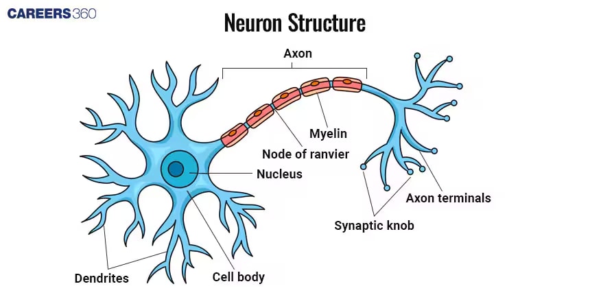
Diagram of Neuron
Given below is the simple neuron diagram with its parts:
Commonly Asked Questions
Structure of a Neuron
The neuron structure contains several primary parts that make the cell functional. These parts include:
Cell Body (Soma)
Contains a nucleus and organelles.
Processes information and maintains cell health.
Dendrites
Branch-like structures which receive signals from other neurons.
Increase surface area for signal reception.
Axon
Long, slender projection which transmits signals away from the cell body.
Axons come in different lengths depending on the type of neuron.
Axon Terminals
These are the endings of the axon that connect to other neurons or muscle cells.
Release neurotransmitters to communicate with other cells.
Myelin Sheath
It is the insulating membrane around axons.
It increases the speed of nerve impulse transmission.
Nodes of Ranvier
Gaps in the myelin sheath
Saltatory conduction quickly transmits the signal.
Commonly Asked Questions
Types of Neurons
Neurons are classified based on the structure and functionality of the neuron. They are classified into:
Sensory Neurons
It carries sensory information from receptors to the central nervous system.
Example: Neuron in the skin which detects touch.
Motor Neurons
Carry commands from the central nervous system to muscles.
Example: Neuron which causes muscle contraction
Interneurons
Connect sensory and motor neurons within the central nervous system.
Example: Neurons in the spinal cord that relay signals between sensory and motor neurons.
Also Read:
| Human Nervous System | Nerve Fibres |
| Autonomic Nervous System | Sympathetic Nervous System |
| Diagram of Eye | Diagram of Ear |
Commonly Asked Questions
Recommended Video on ‘Diagram of Neuron’
Frequently Asked Questions (FAQs)