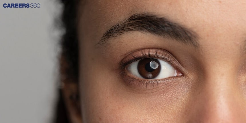
Diagram of Eye: Pictures, Images, Stock Photos

Diagram of Eye: Pictures, Images, Stock Photos

Irshad AnwarUpdated on 02 Jul 2025, 06:48 PM IST

The human eye is a complex organ that enables vision by capturing light and converting it into electrical signals for the brain to interpret. Its structure includes the cornea, lens, retina, iris, and optic nerve, each playing a vital role in the process of sight. In this article, the human eye, external structures, and internal structures are discussed. The human eye is a topic of the chapter Neural Control and Coordination in Biology.

This Story also Contains

  1. Human Eye
  2. External Structures
  3. Internal Structures
Diagram of Eye: Pictures, Images, Stock Photos
Diagram of Eye

Human Eye

An illustrative diagram of the eye is very basic in understanding its anatomy and features. In this way, one learns how the eye works in facilitating vision through the illustration.

External Structures

The extrinsic structures act to protect the eye and facilitate its function.

Eyelids

The eyelids protect the eye from debris and control the amount of light falling on the eye.

Conjunctiva

It is a thin membrane covering the front of the eye and lining the inner eyelids.

Diagram of Human Eye

External structure of Eye

Also Read-

Commonly Asked Questions

Q: What is the function of the sclera in the eye?
A:
The sclera, also known as the "white of the eye," is the tough, fibrous outer layer of the eyeball. Its main functions are: 1) Protecting the eye's internal structures from injury, 2) Maintaining the eye's shape, 3) Providing an attachment point for the extraocular muscles that control eye movement, and 4) Resisting internal and external pressures to keep the eye functioning properly.
Q: What is the function of the conjunctiva in the eye?
A:
The conjunctiva is a clear membrane that covers the white part of the eye (sclera) and the inner surface of the eyelids. Its functions include: 1) Producing mucus to lubricate the eye, 2) Preventing foreign objects from entering behind the eyeball, 3) Providing a protective barrier against pathogens, and 4) Contributing to the immune defense of the eye.
Q: What is the function of the cornea in the eye?
A:
The cornea is the clear, dome-shaped surface at the front of the eye. It serves two primary functions: 1) It acts as a protective barrier for the eye's internal structures, and 2) It refracts (bends) incoming light, providing about 70% of the eye's focusing power before light reaches the lens.
Q: How do tears contribute to eye health and function?
A:
Tears, produced by lacrimal glands, serve multiple purposes: 1) Lubricating the eye to prevent friction during blinking and eye movement, 2) Washing away debris and potential pathogens, 3) Providing oxygen and nutrients to the cornea, 4) Maintaining a smooth optical surface on the cornea for clear vision, and 5) Containing antibacterial enzymes that help protect against infections.
Q: What is the role of melanin in the eye?
A:
Melanin in the eye serves several purposes: 1) In the iris, it determines eye color and helps control the amount of light entering the eye, 2) In the retinal pigment epithelium, it absorbs stray light to improve visual acuity and protect the retina from photo-oxidative damage, 3) It acts as an antioxidant, protecting eye tissues from harmful free radicals, and 4) It helps in the development and maintenance of normal eye structure during embryonic development.

Internal Structures

The structures inside the eye complete the vision.

Cornea

The transparent, curved surface at the front of the eye refracts light.

Iris And Pupil

The iris controls the diameter of the pupil, which regulates the amount of light reaching the eye.

Lens

Flexible, transparent; focusing light onto the retina.

Retina

The inner one with photoreceptor cells which detect the light—rods and cones, which transmit it to the brain.

Internal structure of eye

Also Read-

Recommended video on Diagram of Eye


Commonly Asked Questions

Q: How does the iris control the amount of light entering the eye?
A:
The iris controls light entry by adjusting the size of the pupil. In bright light, the iris muscles contract, making the pupil smaller to reduce light entry. In dim light, the iris muscles relax, enlarging the pupil to allow more light in. This process is called pupillary reflex and helps optimize vision in different lighting conditions.
Q: How does the lens of the eye change shape to focus on objects at different distances?
A:
The lens changes shape through a process called accommodation. When focusing on nearby objects, the ciliary muscles contract, allowing the lens to become more convex (rounded). For distant objects, the ciliary muscles relax, and the lens flattens. This shape change alters the lens's focal length, enabling clear vision at various distances.
Q: What is the blind spot, and why can't we normally perceive it?
A:
The blind spot is the area on the retina where the optic nerve exits the eye, containing no photoreceptors. We don't normally perceive it because: 1) It's small and offset from the center of vision, 2) The brain "fills in" the missing information using surrounding visual data, and 3) Having two eyes compensates for each eye's blind spot.
Q: How do rods and cones differ in their function and distribution on the retina?
A:
Rods and cones are photoreceptor cells with different functions and distributions. Rods are more numerous, sensitive to low light, and responsible for night vision but don't perceive color. They're distributed throughout the retina except in the fovea. Cones are concentrated in the fovea, less sensitive to light, but provide color vision and sharp detail in bright conditions.
Q: What is the macula, and why is it important for vision?
A:
The macula is a small, highly sensitive area near the center of the retina. It's crucial for central vision, providing the sharp, detailed vision needed for activities like reading, recognizing faces, and driving. The macula contains a high concentration of cone cells, making it responsible for our most acute and color-sensitive vision.


Frequently Asked Questions (FAQs)

Q: How does the accommodation reflex work, and what structures are involved?
A:
The accommodation reflex allows the eye to focus on objects at varying distances. It involves: 1) Detection of a blurre
Q: How does the blood-retina barrier function, and why is it important?
A:
The blood-retina barrier is a selective barrier that regulates the passage of substances between the retinal blood vessels and the neural retina. It functions through: 1) Tight junctions between retinal endothelial cells, 2) The retinal pigment epithelium (RPE). Its importance lies in: a) Protecting the retina from potentially harmful substances in the bloodstream, b) Maintaining a controlled environment for proper retinal function, c) Regulating the transport of nutrients and waste products, d) Preventing the entry of immune cells that could damage retinal tissue, and e) Contributing to the immune privilege of the eye.
Q: What is the role of horizontal cells in the retina?
A:
Horizontal cells in the retina are interneurons that play several important roles: 1) They provide lateral inhibition, which enhances contrast and sharpens edges in visual perception, 2) They help regulate the input from photoreceptors to bipolar cells, adjusting sensitivity to different light levels, 3) They contribute to color opponency mechanisms, enhancing color perception, 4) They assist in adapting retinal responses to changes in ambient light, and 5) They help integrate signals from multiple photoreceptors, expanding the receptive fields of other retinal neurons.
Q: How does the eye's refractive system work to focus images on the retina?
A:
The eye's refractive system focuses images on the retina through several components: 1) The cornea provides about 70% of the eye's focusing power by bending light as it enters the eye, 2) The lens fine-tunes the focus through accommodation, changing shape to adjust for viewing distance, 3) The aqueous and vitreous humors provide additional refraction and maintain the eye's shape, 4) The pupil controls the amount of light entering the eye, affecting depth of field, and 5) The overall length of the eye determines where the focal point falls, with emmetropia resulting in a clear image on the retina.
Q: What is the function of bipolar cells in the retina?
A:
Bipolar cells in the retina serve as intermediary neurons between photoreceptors and ganglion cells. Their functions include: 1) Receiving and processing signals from photoreceptors, 2) Transmitting this information to ganglion cells, 3) Performing initial processing of visual information, such as detecting changes in light intensity, 4) Contributing to the center-surround organization of receptive fields, which enhances edge detection and contrast sensitivity, and 5) Participating in parallel processing of different aspects of visual information.
Q: How does the trabecular meshwork contribute to eye health?
A:
The trabecular meshwork is a spongy tissue located near the base of the cornea. Its primary functions are: 1) Regulating the outflow of aqueous humor from the eye, which helps maintain proper intraocular pressure, 2) Acting as a filter to remove cellular debris and other particles from the aqueous humor, 3) Contributing to the immune defense of the eye by trapping pathogens, and 4) Playing a role in the pathogenesis of glaucoma when its function is impaired.
Q: What is the role of the retinal pigment epithelium (RPE) in vision?
A:
The retinal pigment epithelium (RPE) is a layer of cells located between the retina and choroid. Its functions include: 1) Absorbing scattered light to improve visual acuity, 2) Transporting nutrients from the choroid to the retina, 3) Removing waste products from photoreceptor cells, 4) Regenerating visual pigments in photoreceptors, 5) Maintaining the blood-retina barrier, and 6) Phagocytosing shed photoreceptor outer segments, which is crucial for photoreceptor renewal.
Q: What is the purpose of the anterior and posterior chambers in the eye?
A:
The anterior and posterior chambers are fluid-filled spaces in the eye. The anterior chamber lies between the cornea and iris, while the posterior chamber is between the iris and lens. Their functions include: 1) Containing and circulating aqueous humor, which nourishes the cornea and lens, 2) Maintaining intraocular pressure, 3) Providing a clear medium for light to pass through, 4) Facilitating the flow of nutrients and waste products, and 5) Contributing to the overall shape and structure of the eye.
Q: How does the lens capsule contribute to the function of the eye lens?
A:
The lens capsule is a thin, elastic membrane that encapsulates the lens. Its functions include: 1) Maintaining the shape and position of the lens, 2) Providing an attachment point for zonular fibers, which connect the lens to the ciliary body, 3) Acting as a barrier to prevent the passage of large molecules into the lens, 4) Contributing to the process of accommodation by allowing the lens to change shape, and 5) Helping to maintain the transparency of the lens by regulating its internal environment.
Q: What is the function of ganglion cells in the retina?
A:
Ganglion cells are the final output neurons of the retina. Their functions include: 1) Integrating and processing visual information from photoreceptors via bipolar and amacrine cells, 2) Encoding different aspects of visual stimuli such as motion, color, and contrast, 3) Transmitting visual information to the brain through their axons, which form the optic nerve, 4) Contributing to various visual reflexes and circadian rhythm regulation through connections to non-image-forming brain areas.
Articles