Muscles: Types, Groups, Anatomy, Functions, Composition, Development

Muscles: Types, Groups, Anatomy, Functions, Composition, Development

Irshad AnwarUpdated on 02 Jul 2025, 06:45 PM IST

Muscles are specialized tissues responsible for movement and support in the human body. They work by contracting and relaxing, enabling various actions like walking, lifting, and even maintaining posture. In this article, muscles, types of muscles, muscle structure, muscle function, and common muscle disorders and diseases are discussed. Muscles are a topic of the chapter Locomotion And Movement in Biology.

This Story also Contains

  1. What are Muscles?
  2. Types of Muscles
  3. Muscle Structure
  4. Muscle Function
  5. Common Muscle Disorders and Diseases
Muscles: Types, Groups, Anatomy, Functions, Composition, Development
Muscles

What are Muscles?

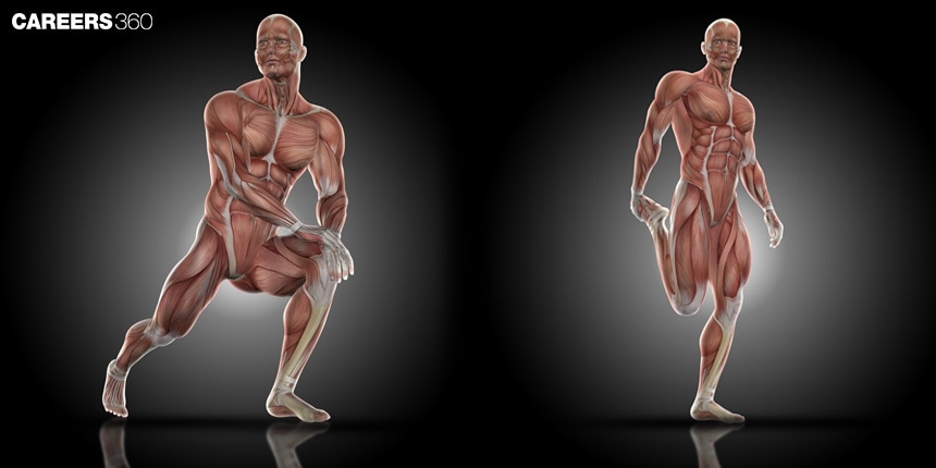
Muscles are specialised tissues in the human body whose major function is to move by their ability to contract. They are essential for a wide range of operations or activities that occur or take place within the body, such as maintaining posture, providing means and enabling locomotion, and assisting in critical functions like breathing and digestion. They contribute to overall health by way of supporting the skeletal system, protecting the internal organs, and offering help in metabolic processes.

Muscles take part in movement but play a crucial role in the overall health of the body. Regular muscle activity improves cardiovascular health, supports metabolic functions, and aids in maintaining a healthy weight. Other than daily movements, muscle strength is also vital for daily acts and the prevention of injuries of any type.
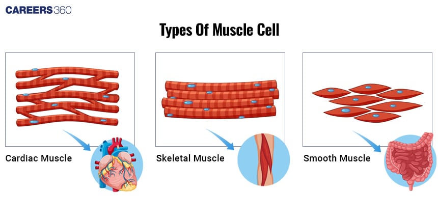
Types of Muscles

The types of muscles in the human body are broadly divided into three categories: skeletal, cardiac, and smooth muscles.

Skeletal Muscles

  • They are striated in appearance.

  • Multinucleated fibers

  • Voluntary control

  • Fixed to bones by tendons

  • Extensively in limbs and torso

  • Cause movement due to contraction and relaxation abilities

  • Examples: Biceps brachii (arm), Quadriceps femoris (thigh)

NEET Highest Scoring Chapters & Topics
Know Most Scoring Concepts in NEET 2024 Based on Previous Year Analysis.
Know More

Cardiac Muscles

  • Striated with intercalated discs

  • One nucleus per cell

  • Involuntary

  • In the Walls of the heart (myocardium)

  • Pump blood throughout the body

  • Contraction occurs continually in a rhythmic manner.

Smooth Muscles

  • Not striated

  • One nucleus per cell

  • Autonomic nervous control

  • In the Walls of hollow organs (e.g., intestine, blood vessels)

  • Move substances along the body

  • Examples: Muscles in the digestive tract, blood vessel walls.

Types of Muscles

Also Read:

Muscle Structure

Muscle structure is complex, comprising different parts that interact to produce contraction and movement.

Muscle Fibers

  • Elongated, cylindrical cells

  • Several nuclei peripherally located

Sarcomere: The Functional Unit

  • Repeating units of myofibrils

  • Units responsible for muscle contraction

Myofibrils, Actin and Myosin Filaments

  • Myofibrils: Contractile threads within muscle fibers

  • Actin (thin) and myosin (thick) filaments: proteins involved in contraction.

Connective Tissue Components

  • Endomysium: surrounds individual muscle fibres

  • Perimysium: encases bundles of fibres (fascicles)

  • Epimysium: encloses the entire muscle

Tendons and their Role in Muscle Attachment:

  • Connect muscles to bones

  • Transmit force from muscle contraction to the skeleton.

Commonly Asked Questions

Q: What is the basic functional unit of a skeletal muscle?
A:
The basic functional unit of a skeletal muscle is called a sarcomere. Sarcomeres are composed of thick and thin filaments (myosin and actin) that slide past each other during muscle contraction, following the sliding filament theory. Multiple sarcomeres form myofibrils, which bundle together to create muscle fibers.
Q: What is the role of titin in muscle structure and function?
A:
Titin is a large protein that spans the length of the sarcomere, connecting the Z-line to the M-line. It plays several important roles: 1) Providing structural support and maintaining sarcomere organization, 2) Contributing to muscle elasticity and passive tension, 3) Serving as a molecular spring during muscle stretching and contraction, and 4) Acting as a scaffold for the assembly of other sarcomeric proteins.
Q: What is the function of the sarcoplasmic reticulum in muscle cells?
A:
The sarcoplasmic reticulum is a specialized form of endoplasmic reticulum in muscle cells. Its primary function is to store and release calcium ions. During muscle contraction, it releases calcium into the sarcoplasm, triggering the sliding of actin and myosin filaments. During relaxation, it actively pumps calcium back into storage, allowing the muscle to relax.
Q: What is the difference between a tendon and a ligament?
A:
Tendons and ligaments are both types of connective tissue, but they serve different functions. Tendons connect muscles to bones, transmitting the force generated by muscle contraction to move the bone. Ligaments, on the other hand, connect bones to other bones, providing stability to joints and limiting excessive movement.
Q: How does skeletal muscle fiber arrangement affect muscle function?
A:
Skeletal muscle fiber arrangement affects the muscle's strength and range of motion. Parallel fibers run the length of the muscle and provide a wide range of motion but less force. Pennate muscles have fibers that attach to the tendon at an angle, allowing more fibers in a given volume, resulting in greater force production but a smaller range of motion.

Muscle Function

The mechanism of muscle function centres on the process of contraction and how muscles contract.

Sliding Filament Theory:

  • Actin and myosin filaments slide past each other

  • Shortens the sarcomere, producing contraction.

Role of ATP and Calcium Ions:

  • ATP provides energy for contraction

  • Calcium ions control the interaction of actin and myosin.

Neuromuscular Junction and Action Potential:

  • Synapse between a motor neuron and muscle fibre.

  • Action potential leads to muscle contraction.

Isotonic and Isometric Contractions:

  • Isotonic: length of the muscle changes (e.g. lifting a weight)

  • Isometric: muscle does not change in length (e.g. holding a position)

Concentric and Eccentric Contractions:

  • Concentric: Muscle shortens when contracting

  • Eccentric: Muscle lengthens when contracting

Commonly Asked Questions

Q: How does the sliding filament theory explain muscle contraction?
A:
The sliding filament theory explains that muscle contraction occurs when myosin heads attach to actin filaments and pull them towards the center of the sarcomere. This causes the sarcomere to shorten without the filaments themselves changing length. As multiple sarcomeres shorten simultaneously, the entire muscle contracts.
Q: What is the role of calcium in muscle contraction?
A:
Calcium plays a crucial role in muscle contraction by binding to troponin, a regulatory protein on actin filaments. This binding causes a conformational change that moves tropomyosin away from the myosin-binding sites on actin. As a result, myosin can attach to actin, initiating the power stroke and muscle contraction.
Q: What is the difference between isotonic and isometric muscle contractions?
A:
Isotonic contractions involve the muscle changing length while maintaining relatively constant tension. This occurs when lifting or lowering a weight. Isometric contractions, on the other hand, involve the muscle generating force without changing length, such as when pushing against an immovable object or holding a static position.
Q: What is the function of the neuromuscular junction?
A:
The neuromuscular junction is the synapse between a motor neuron and a muscle fiber. It serves as the site where nerve impulses are transmitted to the muscle, triggering contraction. When an action potential reaches the neuromuscular junction, it causes the release of acetylcholine, which binds to receptors on the muscle fiber, initiating the contraction process.
Q: How does muscle tone differ from muscle contraction?
A:
Muscle tone refers to the continuous, partial contraction of muscles even when at rest. It maintains posture and readiness for action. Muscle contraction, in contrast, is the active shortening of muscle fibers in response to stimulation, resulting in movement or force generation. Tone is maintained by low-level, continuous nerve impulses, while contraction requires stronger, specific nerve signals.

Common Muscle Disorders and Diseases

Several disorders and diseases can compromise muscle and thus function and quality of life.

Muscular Dystrophy

  • Duchenne muscular dystrophy: Progressive muscle weakness

  • Becker muscular dystrophy: Similar but milder

Genetic Basis and Treatment Options:

  • Genetic mutations that alter muscle proteins.

  • Treatment is symptomatic and aims to retard the progression.

Myasthenia Gravis

  • Muscle weakness, fatigue

  • Autoimmune disorder at the neuromuscular junction.

Treatment and Management:

  • Medications aimed at improving nerve-muscle communication.

  • Immunosuppressive therapies.

Muscle Cramps and Strains

  • It is caused by dehydration, overuse, and electrolyte imbalance.

  • This can be prevented by regular stretching and proper hydration.

Treatment and First Aid:

  • Rest, application of ice, compression, elevation (RICE)

  • Gentle stretch and rehydrate

Also Read:


Recommended Video on Muscles


Frequently Asked Questions (FAQs)

Q: How does the all-or-none principle apply to muscle fibers?
A:
The all-or-none principle states that a muscle fiber either contracts fully or not at all in response to a stimulus. If the stimulus is below the threshold, no contraction occurs. Once the threshold is reached, the muscle fiber contracts with full force. The strength of muscle contraction is regulated by the number of fibers recruited, not by varying the strength of individual fiber contractions.
Q: What is the role of tropomyosin and troponin in muscle contraction?
A:
Tropomyosin and troponin work together to regulate muscle contraction. Tropomyosin is a protein that wraps around the actin filament, covering the myosin-binding sites. Troponin is a complex of three proteins (T, I, and C) attached to tropomyosin. When calcium binds to troponin C, it causes a conformational change that moves tropomyosin away from the myosin-binding sites on actin, allowing myosin to attach and initiate contraction.
Q: How does muscle fiber type distribution affect athletic performance?
A:
Muscle fiber type distribution significantly influences athletic performance. Athletes in power and sprint events tend to have a higher proportion of fast-twitch fibers, allowing for explosive, short-duration efforts. Endurance athletes often have a higher percentage of slow-twitch fibers, supporting sustained, lower-intensity activities. While genetics play a role in fiber type distribution, training can influence fiber characteristics to some extent, optimizing performance for specific activities.
Q: What is the role of creatine phosphate in muscle energy metabolism?
A:
Creatine phosphate serves as a rapid energy source in muscle cells. It can quickly regenerate ATP from ADP by transferring its phosphate group, a reaction catalyzed by creatine kinase. This system is crucial for high-intensity, short-duration activities, providing energy faster than glycolysis or oxidative phosphorylation. It helps maintain ATP levels during the initial seconds of intense exercise and during rapid transitions between rest and activity.
Q: How do cardiac muscle cells differ from skeletal muscle cells?
A:
Cardiac muscle cells differ from skeletal muscle cells in several ways: 1) They are branched and interconnected, forming a functional syncytium, 2) They have intercalated discs that allow for coordinated contraction, 3) They contain more mitochondria, supporting continuous aerobic metabolism, 4) They can generate their own rhythmic contractions (autorhythmicity), 5) They are typically uninucleated but larger than skeletal muscle cells, and 6) They are resistant to fatigue and can function continuously throughout life.
Q: What is the role of myosin light chains in muscle contraction?
A:
Myosin light chains are smaller protein subunits associated with the myosin heavy chains in muscle fibers. They play several roles: 1) Structural support for the myosin molecule, 2) Regulation of myosin ATPase activity, influencing contraction speed and force, 3) Modulation of calcium sensitivity in smooth muscle contraction, and 4) Involvement in the fine-tuning of muscle contraction through phosphorylation, which can affect the rate of cross-bridge cycling.
Q: What is the difference between fast glycolytic and fast oxidative muscle fibers?
A:
Fast glycolytic (FG) and fast oxidative (FOG) fibers are both types of fast-twitch muscle fibers, but they differ in their metabolic properties: 1) FG fibers rely primarily on anaerobic glycolysis for energy, while FOG fibers have a higher capacity for oxidative metabolism, 2) FOG fibers have more mitochondria and myoglobin than FG fibers, 3) FOG fibers are more resistant to fatigue than FG fibers, 4) FG fibers are better suited for short, intense bursts of activity, while FOG fibers can sustain activity for longer periods.