Simple Microscope - Definition, Diagram, FAQs

Simple Microscope - Definition, Diagram, FAQs

Edited By Vishal kumar | Updated on Jul 02, 2025 04:35 PM IST

Introduction of a Microscope: How to make a microscope?

What is a Simple Microscope?
Simple Microscope Definition: A Simple Microscope meaning is used to see a magnified image of an object. Antonie Van Leeuwenhoek, a Dutchman, invented the first simple microscope, consisting of a single powerful magnetic lens that rotates to detect tiny freshwater insects. It is composed mainly of light microscopes. The main property of the target lens used in microscopes is to produce a virtual, upright, and magnified image when the object is placed within a fixed height. A convex lens used in a microscope is to create a simple microscope. Convelenses are widely used as a reading glass or magnifying glass. Now, to obtain high magnification, a combination of two or more convex lenses is used to create an integrated microscope.

Simple Microscope - Definition, Diagram, FAQs

What Are the Parts of a Simple Microscope?

A Simple Microscope contains various optical components and other supporting (or mechanical) components as discussed in this section, the layout of a simple basic microscope with its various labeled numbers. The Eyepiece is connected to the lens via a tube. The eyeball is the lens where the image of the object can be seen. Tube length varies by rotating the button so that a clear image is obtained by changing the focus. The objective lens enhances magnification.

Also read -

A simple Schematic of the Magnifying power of a compound microscope showing its different parts.

Parts of a Simple Microscope

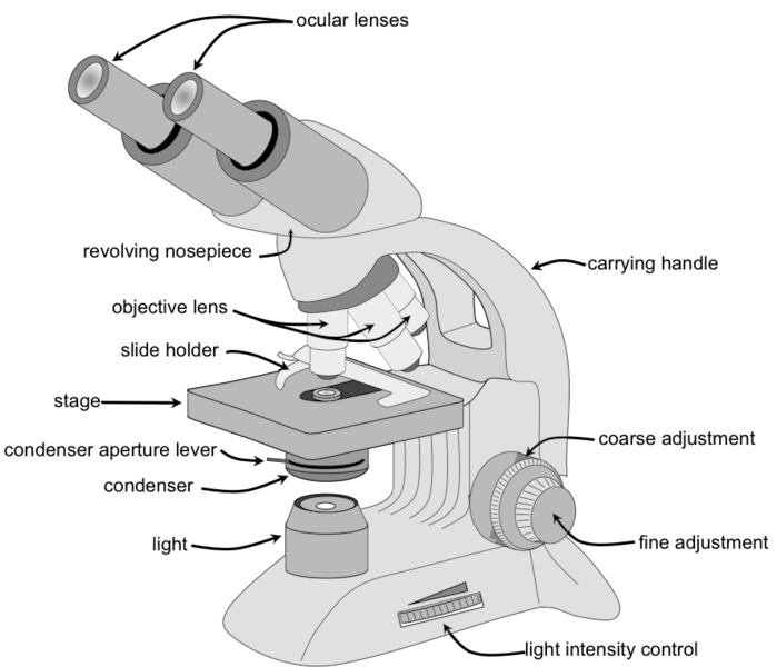
The sample platform, made of metal foil, consists of metal clips holding a sample, which is placed on a glass slide, under observation. The mirror focuses on the light of the sample. All components are grounded.

The base is a component of equipment that provides support for the capture of other parts of the microscope. The arm of the microscope is connected to a physical object.

Lens used in simple microscope magnification

Basically, increasing the size of the object and the size of the image. The lens magnification figure is given below:

Magnification (M) = Himage / Object

However, in the case of a simple microscope, a short-lived convex lens is used to magnify the image of an object, and the angle is passed to the eye for object and image.

The distance between the object and the center of the curved lens.

Using a lens formula given as :

$\frac{1}{F}=\frac{1}{-U}-\frac{1}{-D}$

, the number 3 can be written as:

$M=1+\frac{D}{F}$

Here, M describes the magnification of a simple microscope, D is a small distance of contrast view and F is the focal length of the convex lens. Since ‘F’ is in the denominator in the equation of magnification, that is why a short length of focus can lead to higher magnification.

A Simple Microscope trial

Here, we perform a simple test to calculate the magnitude of an F-centered biconvex lens that acts as a magnifying glass.

Resources needed:

Biconvex lens with short length focusing on ‘F’ and handle.

Newspaper article with low font size.

Process:

Place the reading under the biconvex lens with your eyes next to the lens.

Slowly move the lens to the article and move your head through the lens to view it.

As the lens moves closer to the article, you will notice that the letters of the alphabet will become smaller and blurry in reading.

As you move towards the lens you will see a clear and enlarged image of the printed alphabet in the articles. Think of a position as an 'A'. In this position, let's say the distance between the lens and the article is 'D'.

If you pass this position, the image will be enlarged but blurred and difficult to read.

Using the formula, $M=1+\frac{D}{F}$

magnification can be calculated.

NEET Highest Scoring Chapters & Topics
This ebook serves as a valuable study guide for NEET exams, specifically designed to assist students in light of recent changes and the removal of certain topics from the NEET exam.
Download E-book

What Are the Parts of a Simple Microscope?

The following are the components of a Simple Microscope and their functions:

Eyeball: A lens used to study samples and placed on top. It has an increase of 10X to 15X.

Baseline: This provides microscope support.

Tube: This is used to connect an eyepiece to the target lens.

Target lenses: These are available in 10X, 40X, and 100X magnification lenses and have color coding. The lower electric lenses are shorter lenses and the upper electric lenses are much longer.

A flexible nose piece: This is also known as a turret. It is used to capture other purpose lenses and can be changed while viewing samples.

Diaphragm: Used to control the amount of light passing through the stage.

Stage: It is a platform used to place slides with samples.

Stage Clip: This is used to hold slides in the right place.

Adjust location button: Used to focus on scanning.

Good repair box: Used to focus on oil.

Arm: Used to support the tube and connect to the base of the microscope.

Power switch: A primary power switch used to turn the microscope on or off.

Condenser: Used to focus the light on the sample and uses 400X electric lenses.
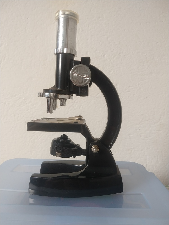
 Simple Microscope

Simple Microscope

Use of a Simple Microscope

  • Used in pedology (study of soil particles)
  • It is used by a dermatologist to diagnose various skin diseases.
  • It is used in microbiology to study algae samples, fungi etc.
  • It is used by carpenters to obtain an enlarged view of fine jewelry.

Also check-

NCERT Physics Notes:

Frequently Asked Questions (FAQs)

1. How many lenses are in a simple microscope?

 A Simple Microscope contains a single lens that is traditionally called a loupe. A well-known example today is the reading or magnifying glass. Modern high-resolution lenses are usually made of two-dimensional glass materials that produce a colour-coded image.

2. Which lens is used in a simple microscope?

A convex lens is used to create a simple microscope. A convex lens is widely used and widely used as a reading glass or magnifying glass.

3. What is an example of a simple microscope?

Examples of simple microscopes include reading glasses, decorative ornaments, and pocket magnifiers. ... There are two types of integrated microscopes that use light to detect visible (or flexible) objects and invisible objects. An opaque object requires reflective light from an object through the optic tube lens.

4. Which convex lens is used with a simple microscope?

Converting the lens is called a convex lens that converts the rays of the light of the event to the same as the main axis at the same time; this is why flexible lenses are used in objects such as microscopes to focus on minute particles.

5. Why is a convex lens used with a microscope?

In the microscope, we often use a convex lens because the convex lens even magnifies images. Microscopes produce highly enlarged images of very small objects for this purpose the convex app is very useful. ... However, between the three lenses, the lens located at the end of the microscope produces a transformed and enlarged image.

6. What is a simple microscope?
A simple microscope, also known as a magnifying glass, is an optical instrument that uses a single convex lens to produce a magnified image of a small object. It works by placing the object within the focal length of the lens, creating an enlarged, virtual, and erect image.
7. How does a simple microscope differ from a compound microscope?
A simple microscope uses only one lens for magnification, while a compound microscope uses multiple lenses. Simple microscopes provide lower magnification (typically up to 10x) but are more portable and easier to use. Compound microscopes offer higher magnification and resolution but are more complex.
8. What is the principle behind the magnification in a simple microscope?
The magnification in a simple microscope is based on the principle of refraction. When an object is placed within the focal length of a convex lens, light rays from the object are refracted to form a virtual, enlarged image on the same side of the lens as the object.
9. Can a simple microscope produce a real image?
No, a simple microscope cannot produce a real image. It always forms a virtual image because the object is placed within the focal length of the lens. The image formed is erect, enlarged, and on the same side of the lens as the object.
10. What determines the magnification power of a simple microscope?
The magnification power of a simple microscope depends on two factors: the focal length of the lens and the distance between the object and the lens. A shorter focal length and a smaller object distance result in higher magnification.
11. Can a concave lens be used as a simple microscope?
No, a concave lens cannot be used as a simple microscope. Concave lenses always produce diminished virtual images, regardless of object position. Simple microscopes require magnification, which is achieved using convex lenses.
12. How does the position of the object affect the image in a simple microscope?
The object must be placed within the focal length of the lens in a simple microscope. As the object moves closer to the focal point, the magnification increases, and the virtual image appears farther away. If the object is placed at or beyond the focal point, no useful image is formed.
13. What is the significance of the near point in relation to a simple microscope?
The near point is the closest distance at which the human eye can focus comfortably (typically 25 cm for a normal eye). The magnification of a simple microscope is often defined in relation to this near point, as it determines how much larger the image appears compared to viewing the object unaided at the near point.
14. How is angular magnification defined for a simple microscope?
Angular magnification for a simple microscope is defined as the ratio of the angle subtended by the image at the eye to the angle subtended by the object when placed at the near point. It is given by the formula: M = 1 + D/f, where D is the near point distance and f is the focal length of the lens.
15. What are the advantages of a simple microscope over a compound microscope?
Simple microscopes are more portable, easier to use, and less expensive than compound microscopes. They also provide a wider field of view and are useful for quick observations of larger specimens. Additionally, simple microscopes produce erect images, making them easier to interpret.
16. What is the difference between linear and angular magnification in a simple microscope?
Linear magnification refers to the ratio of image size to object size, while angular magnification is the ratio of the angle subtended by the image to the angle subtended by the object at the near point. In simple microscopes, angular magnification is more relevant as it relates directly to how much larger the image appears to the eye.
17. Can a simple microscope be used for measuring the size of objects?
While a simple microscope can provide a magnified view of an object, it is not inherently designed for precise measurements. However, if the microscope is equipped with a calibrated scale in the eyepiece or on the stage, it can be used for approximate size estimations. For more accurate measurements, specialized measuring microscopes or digital imaging systems are preferred.
18. How does spherical aberration affect the image quality in a simple microscope?
Spherical aberration in a simple microscope causes light rays passing through different parts of the lens to focus at slightly different points. This results in a slightly blurred or distorted image, especially near the edges. The effect becomes more pronounced with higher magnifications and can reduce the overall clarity and sharpness of the image.
19. How does the concept of depth of field apply to simple microscopes?
Depth of field in a simple microscope refers to the range of distances within which objects appear acceptably sharp. Simple microscopes generally have a greater depth of field compared to high-power compound microscopes. This means that a larger portion of the object remains in focus at once, which can be advantageous for observing three-dimensional specimens.
20. Can a simple microscope be used for photomicrography?
While it's possible to take photographs through a simple microscope, it's not ideal for photomicrography. The virtual image formed by a simple microscope cannot be projected onto a screen or sensor. However, with careful setup and proper focusing, it's possible to capture images using a camera positioned where the eye would normally be, though the results may not be as high-quality as those obtained with compound microscopes designed for photomicrography.
21. What is the role of the iris of the eye when using a simple microscope?
The iris of the eye plays a crucial role when using a simple microscope by controlling the amount of light entering the eye. It adjusts the pupil size to optimize the light level for the magnified image, helping to balance brightness and image clarity. This adaptation is particularly important because the light intensity can vary significantly depending on the magnification and the object being viewed.
22. How does the simple microscope demonstrate the concept of linear magnification?
While angular magnification is more commonly used for simple microscopes, they also demonstrate linear magnification. Linear magnification is the ratio of image size to object size. In a simple microscope, the virtual image is always larger than the object, showing positive linear magnification. This can be calculated using the formula m = v/u, where v is the image distance and u is the object distance.
23. Why is the image formed by a simple microscope virtual?
The image formed by a simple microscope is virtual because the refracted light rays do not actually meet but appear to diverge from a point behind the lens. Our eyes perceive these diverging rays as coming from an enlarged, upright image.
24. What is the relationship between the focal length of the lens and the magnification in a simple microscope?
There is an inverse relationship between the focal length and magnification in a simple microscope. A shorter focal length results in higher magnification. The magnification is approximately equal to the near point distance (25 cm) divided by the focal length of the lens.
25. How does the simple microscope relate to the human eye?
The simple microscope assists the human eye by creating an enlarged virtual image that subtends a larger angle at the eye than the object would at the near point. This allows the eye to perceive more detail than it could when viewing the object directly.
26. Can a simple microscope correct for visual defects like myopia or hypermetropia?
While a simple microscope is not designed to correct visual defects, it can sometimes help people with certain visual impairments see small objects more clearly. However, for proper correction of myopia or hypermetropia, prescription eyeglasses or contact lenses are necessary.
27. How does the working of a magnifying glass relate to the simple microscope?
A magnifying glass is essentially the same as a simple microscope. Both use a single convex lens to produce an enlarged, virtual image of an object placed within the focal length of the lens. The principles of operation and image formation are identical.
28. What happens to the image if the object is placed exactly at the focal point of a simple microscope?
If the object is placed exactly at the focal point of the lens in a simple microscope, no useful image is formed. The refracted rays would emerge parallel to each other, and the image would appear to be at infinity, making it impossible to view.
29. How does the aperture of the lens affect the performance of a simple microscope?
The aperture of the lens affects the amount of light gathered and the resolution of the image. A larger aperture allows more light to enter, producing a brighter image, and can potentially improve resolution. However, it may also introduce aberrations if not properly designed.
30. Can a simple microscope produce a magnification greater than 100x?
While theoretically possible, it is impractical for a simple microscope to produce magnifications greater than about 10x. Higher magnifications would require extremely short focal lengths, leading to very small working distances and significant aberrations. For higher magnifications, compound microscopes are more suitable.
31. How does chromatic aberration affect the image in a simple microscope?
Chromatic aberration in a simple microscope causes different colors to focus at slightly different points, resulting in colored fringes around the edges of the image. This effect is more noticeable with higher magnifications and can reduce image clarity and contrast.
32. What is the working distance in a simple microscope, and why is it important?
The working distance in a simple microscope is the space between the lens and the object being viewed. It's important because it determines how close you need to get to the object for proper focus. Shorter focal length lenses provide higher magnification but also reduce the working distance, making it more challenging to manipulate or illuminate the specimen.
33. How does the field of view change with increasing magnification in a simple microscope?
As the magnification increases in a simple microscope, the field of view decreases. This means that at higher magnifications, you can see more detail but in a smaller area of the specimen. This trade-off between magnification and field of view is an important consideration when using simple microscopes.
34. How does the quality of the lens affect the performance of a simple microscope?
The quality of the lens significantly impacts the performance of a simple microscope. Higher quality lenses with better optical properties (such as reduced aberrations and improved coating) provide clearer, sharper images with less distortion. They also typically offer better color rendition and can achieve higher useful magnifications.
35. What is the role of the eye in the functioning of a simple microscope?
The eye plays a crucial role in the functioning of a simple microscope. It acts as the final optical element in the system, focusing the virtual image produced by the lens onto the retina. The brain then interprets this image, perceiving it as larger than the actual object. The simple microscope relies on the eye's ability to accommodate and focus on the virtual image at various distances.
36. How does the simple microscope demonstrate the concept of angular magnification?
The simple microscope demonstrates angular magnification by increasing the angle subtended by the object at the eye. When an object is viewed through the microscope, it appears larger because the virtual image subtends a larger angle at the eye than the object would if viewed at the near point. This increase in angular size is what allows us to perceive more detail.
37. Can a simple microscope be used to view microscopic organisms like bacteria?
While a simple microscope can provide some magnification, it is generally not powerful enough to view most microscopic organisms like bacteria. Simple microscopes typically offer magnifications up to about 10x, which is insufficient for observing microorganisms. For viewing bacteria and other microorganisms, compound microscopes with much higher magnification capabilities are necessary.
38. How does the simple microscope relate to Snell's law of refraction?
Snell's law of refraction is fundamental to the operation of a simple microscope. As light passes from the object through the lens, it bends according to Snell's law. This bending of light rays is what allows the lens to form a magnified virtual image. The degree of bending depends on the refractive indices of the lens material and the surrounding medium, as described by Snell's law.
39. What is the difference between the magnification and the resolving power of a simple microscope?
Magnification refers to how much larger an object appears when viewed through the microscope, while resolving power is the ability to distinguish between two closely spaced points as separate entities. A simple microscope may provide significant magnification, but its resolving power is limited compared to more advanced microscopes. Increasing magnification beyond the limits of resolving power results in an enlarged but blurry image.
40. How does the curvature of the lens affect the performance of a simple microscope?
The curvature of the lens directly affects its focal length and, consequently, its magnifying power. A more steeply curved lens (with a smaller radius of curvature) has a shorter focal length and provides higher magnification. However, increased curvature can also lead to more pronounced optical aberrations, potentially reducing image quality if not properly designed.
41. Can a simple microscope produce a magnified inverted image?
No, a simple microscope cannot produce a magnified inverted image. It always forms an erect (upright) virtual image. Inverted images in microscopy are typically associated with compound microscopes, where multiple lenses are used to form real, inverted images.
42. How does the refractive index of the lens material affect the performance of a simple microscope?
The refractive index of the lens material affects the bending of light rays and thus the focal length of the lens. A higher refractive index material can produce a shorter focal length for the same lens curvature, resulting in higher magnification. However, higher refractive index materials may also introduce more chromatic aberration if not properly designed.
43. What is the significance of the lens equation in understanding simple microscopes?
The lens equation (1/f = 1/u + 1/v, where f is focal length, u is object distance, and v is image distance) is crucial for understanding simple microscopes. It helps in calculating the position and size of the virtual image formed. For simple microscopes, the object distance (u) is always less than the focal length (f), resulting in a negative value for the image distance (v), indicating a virtual image.
44. Can a simple microscope be used for observing opaque objects?
Yes, a simple microscope can be used to observe opaque objects, but it requires proper illumination. Unlike transparent specimens that can be illuminated from below, opaque objects need top illumination. Many simple microscopes or magnifying glasses are designed with built-in lights or can be used with external light sources to effectively view opaque objects.
45. What is the relationship between the object distance and image distance in a simple microscope?
In a simple microscope, the object is always placed within the focal length of the lens. As a result, the image distance is always negative (indicating a virtual image) and on the same side of the lens as the object. As the object moves closer to the lens, the virtual image appears to move further away and becomes larger.
46. How does the simple microscope demonstrate the principle of reversibility of light?
The principle of reversibility of light states that light follows the same path whether it travels from object to image or vice versa. In a simple microscope, if we were to place a light source at the position of the virtual image, the light rays would travel back through the lens and converge at the object's position, demonstrating this principle.
47. How does the simple microscope relate to the concept of visual angle?
The simple microscope increases the visual angle subtended by an object at the eye. The visual angle is the angle formed by light rays from the extremities of an object at the eye. By creating a larger virtual image, the simple microscope increases this angle, making the object appear larger and allowing us to see more detail than we could with the unaided eye.
48. Can a simple microscope be used to view objects underwater?
A simple microscope can be used to view objects underwater, but it requires some modifications. The refractive index of water is different from that of air, which affects the focal length of the lens. Specialized waterproof magnifiers or underwater microscopes are designed to account for this change in refractive index and provide clear magnification in aquatic environments.
49. How does the simple microscope relate to the concept of optical power?
The optical power of a lens, measured in diopters, is the reciprocal of its focal length in meters. In a simple microscope, a lens with higher optical power (shorter focal length) provides greater magnification. The concept of optical power is directly related to the magnifying capability of the simple microscope, with higher power lenses offering stronger magnification.
50. What is the effect of using multiple simple microscopes in series?
Using multiple simple microscopes in series effectively creates a compound microscope. Each lens in the series further magnifies the image produced by the previous lens. However, this also increases complexity, reduces the field of view, and can introduce more aberrations. While possible, it's generally more practical to use a properly designed compound microscope for higher magnifications.
51. How does the simple microscope demonstrate the principle of minimum deviation?
The principle of minimum deviation states that light passing through a prism experiences the least bending when it enters and exits the prism symmetrically. While not directly applicable to simple microscopes, this principle is related to the optimal design of lenses. In a well-designed simple

Articles

Back to top